A 60-something man without significant past history presented after sudden onset of aphasia 45 minutes prior. There was also a report of a few days of abdominal pain, nausea and vomiting, and of not being himself.
BP was 165/80, pulse 110. He had profound expressive aphasia but his motor exam was intact.
A stroke code was called and the patient underwent a CT stroke series. Non-contrast scan showed no bleed and no evidence of infarct. Cerebral CT angiography did not show a large vessel occlusion.
He was rapidly given tPA (alteplase).
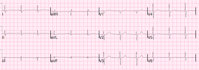
An ECG was obtained (ECG-1):
A previous was available from 6 years prior that was normal:
ECG-1: There is a QS-wave in V2, with ST Elevation and some T-wave inversion. There is a tiny r-wave in V3, also with STE and TWI. There is STE in V4 and V5, with some T-wave inversion. Is this LV aneurysm? Or acute MI? Or subacute MI?
If there is a single lead in V1-V4 which has a T/QRS ratio > 0.36, then there is likely acute MI. If the ratio is <0.36, it is likely old (LV aneurysm) or subacute; symptoms of >6 hours duration suggest subacute, and <6 hours as acute. The ratio in lead V2 is 3.5/5.0 = 0.70, so this is an acute anterior MI. The QS-wave suggests that it is nearly complete. It could also be an old MI with superimposed acute LAD occlusion.
This is diagnostic of Anterior MI, acute or subacute, but not simply an old anterior MI with persistent STE (not just an LV aneurysm -- there is acute ischemia). Acute transmural anterior MI puts patients at high risk of LV mural thrombus, which then can release thrombi and cause stroke or other arterial emboli.
I remember the time in the early 1980s, when I was in medical school, when few patients received reperfusion therapy for STEMI/OMI, and thus most anterior MI progressed to completion (transmural) if they did not spontaneously reperfuse. All of these patients were put on heparin to prevent mural thrombus.
The first hs troponin I returned at 8000 ng/L, confirming subacute anterior MI.
The patient went emergently to the cath lab and was found to have a 99% acute LAD stenosis with thrombus and TIMI III flow. (Was it open because of the tPA?) It was stented. There was mild CAD in the RCA and LCx.
An echocardiogram showed a wall motion abnormality, and EF of 35%, consistent with a large LAD infarct and it showed a "possible" left ventricular mural thrombus.
Cardiac MRI:
Delayed enhancement sequences obtained at 10 mins after gadolinium administration reveal extensive and patchy subendocardial enhancement involving the mid to distal anterior wall and anteroseptal wall involving less than 50% thickness. There is also delayed enhancement in the apex, involving more than 50% of myocardial thickness.
Impression:
1) Moderately decreased LV function (calculated ejection fraction of 33%) with large wall motion abnormality in the LAD territory.
2) No evidence for left ventricular thrombus. Large area of microvascular obstruction in the apex.
3) Large area of subendocardial delayed enhancement in the mid to distal anterior wall and distal septum, involving less than 50% of myocardial thickness. The LV apex shows transmural enhancement. These findings suggest apart from the apex the rest of the LAD territory is viable
Clinical Course:
The patient had an uneventful hospital course, was transferred to rehab, and later found dead in his bed shortly before discharge.
This illustrates that one of the longer term complications of myocardial infarction is sudden death, and it is not always predictable. (However, the topic of preventing sudden death after myocardial infarction is a big one and beyond the scope of this blog post). An autopsy was inconclusive, but did show "transmural infarct of the anterior and lateral walls," contradicting the MRI results, but consistent with the ECG.
There was also some stenosis distal to the stent, and 80-85% RCA stenosis and 65-70% LCx stenosis. The autopsy results of the coronaries is different from the angiogram results. Why? This probably reflects the problem with autopsy sections of coronary arteries: Because sections can reveal a large amount of extraluminal atherosclerosis, the stenosis appears to be much greater on pathologic section than on angiogram, which tells you only about the lumen of the artery, not the extraluminal plaque..
I suspect that the patient had an arrhythmic death.
Learning Points:
1. An ECG is helpful in identifying the etiology of acute stroke. If there is completed anterior MI, LV aneurysm, or atrial fibrillation, the etiology is likely to be embolic.
2. Completed anterior MI results in a profound wall motion abnormality. The resulting "stasis," combined with inflammation of infarction, creates a perfect environment for an LV mural thrombus, which puts the patient at risk for systemic emboli, including stroke and mesenteric ischemia.
3. Beware the risk of sudden arrhythmic death after acute MI, even when the artery has been stented and the acute ischemia has resolved. That infarcted myocardium is a nidus of arrhythmia.
4. tPA is not a contraindication to coronary angiography (in fact, tPA at a referral hospital before transfer to a PCI institution is recommended if first medical contact to PCI time will be greater than 120 minutes)
5. Myocardial infarction may have very nonspecific symptoms. For this patient, they were abdominal pain, "not being himself," and nausea and vomiting.
6. Autopsy coronary stenosis is usually much greater than seen on angiography


No comments:
Post a Comment
DEAR READER: I have loved receiving your comments, but I am no longer able to moderate them. Since the vast majority are SPAM, I need to moderate them all. Therefore, comments will rarely be published any more. So Sorry.