Saturday, July 30, 2016

Symptomatic Bradycardia. So-called Trifascicular Block. Occum's Razor and Hickum's dictum.

A middle-aged male with PMH significant for atrial fibrillation, cocaine use, DM, HTN, hyperlipidemia, and previous MI that was related to cocaine presented for 5 days of intermittent left-sided chest pain.  He reports that the pain is 10/10, sharp, non-radiating, and lasts for "a couple seconds," is associated with SOB and diaphoresis, and then dissipates/resolves on it's own.  Pain does not change with activity or position.   

Here is his initial ECG:
What do you see?


There is an irregular sinus bradycardia and both bifascicular block (RBBB and LAFB) and 1st degree AV block with a PR interval of 260 ms.  The last complex comes so late that there is a junctional escape before the P-wave can conduct.  However, every P-wave which gets the chance to conduct, does conduct.  There is no evidence of complete AV block.

 Bifascicular block + 1st degree AV block is inappropriately called "trifascicular block," a known misnomer.

Why is it a misnomer?  There is AV block and bifascicular block, but there are NOT 3 blocked fascicles as "tri-" implies.  If the right bundle, left anterior fascicle AND left posterior fascicle were all 3 blocked, that would be true trifascicular block and would be, by definition, complete ("third degree") AV block at the infra-HIS location.

However, it is true that when there is 1st degree AV block in addition to bifascicular block, it makes the AV block more likely to be below the bundle of HIS and makes subsequent complete heart block more likely.

Remember that RBBB and LAFB in the setting of STEMI implies a huge MI (from LAD or left main occlusion and is very dangerous.

Whenever there is high grade block and/or bradycardia, one must think of hyperkalemia and ischemia, and of course medications such as beta blockers and calcium channel blockers, although these latter will mostly cause sinus bradycardia and/or AV block, not block of the bundle of HIS.


The K returned, = 4.5 mEq/L.   Troponins were negative.   Here is the old ECG from 4 years prior:
"So-called" Trifascicular block was present then, but without the sinus bradycardia


This was recorded 22 minutes after the first:
There is profound sinus bradycardia, so much so that complexes 3, 4, and 7 are junctional escapes.


Overnight, it was reported that the patient had episodes of third degree AV block.

Here is the only 12-lead that was recorded:
There are no P-waves at all.  
One cannot call it sinus arrest for certain, because there is an escape at a rate of 38
(sinus arrest requires at least a 2 second pause, some say 3 seconds).

Such an escape is a bit too slow for a junctional escape, which generally happens at 40-60.

Furthermore, a junctional escape should have the same morphology as a conducted sinus rhythm.

However, this morphology is similar to RBBB with Posterior fascicular block, which implies an escape near the anterior fascicle.

Bizarre T-wave inversions (see link below)


Therefore, this is a ventricular escape rhythm.  The bizarre T-wave inversions are common in this situation. See this post:

Bizarre T-wave inversion of Stokes Adams attack (syncope and complete AV block), with alternating RBBB and LBBB


There is no evidence of third degree AV block here.

So this patient has 2 issues:

1) Complete heart block was seen on his monitor, but not recorded on a 12-lead
2) Long sinus pauses (which could be sinus arrest or simply sinus bradycardia, but since the escape happens before a 2-3 second pause can complete, one cannot be certain).

He did get an implanted pacemaker.


"Trifascicular" Block

For the reasons stated above, the 2009 AHA/ACCF/HRS scientific statement on the standardization and interpretation of the electrocardiogram recommends against using the term trifascicular block. 

Chronic bifascicular block in an asymptomatic individual (as this patient had 4 years prior) is associated with a low risk of progression to complete heart block. In contrast, a new bifascicular block with acute anterior myocardial infarction carries a much greater risk of complete heart block. 


Alternating right and left bundle branch block is interpreted as a sign of trifascicular block. In contrast, first degree AV block plus bifascicular block does notnecessarily indicate trifascicular involvement, since this combination can reflect slow conduction in the AV node with concomitant bifascicular block.


This 1981 Article from Circulation (volume 64(6):1265 (full text), studied patients with chronic asymptomatic bifascicular block (as in our patient above).  N = 329 with RBBB and LAFB, 46 with RBBB and LPFB, and 142 patients with LBBB.  These patients were divided, based on EP studies, into those with a prolonged (
greater than or equal to 56 ms) HIS-ventricle (HV) interval ( (n=319) vs. those with a normal HV interval (less than or equal to 55 ms, n = 198).   [A prolonged HV interval would result in a prolonged PR interval and the misnomer "trifascicular block" would be applied to these patients.]  Patients with prolonged HV interval were more likely to have evidence of organic heart disease such as cardiomegaly, CHF, PVCs, and angina.  Over a follow up period averaging 3.7 years, the development of true "trifascicular block," as defined by 2nd or 3rd degree AV block below the bundle of HIS, was 0.6% in the normal HV conduction group and 4.5% in the prolonged group.  


Thus, these patients with chronic asymptomatic bifascicular block and 1st degree AV block may have infra-HIS conduction delay (not just AV node delay).  Of course you do not know in the ED whether the delay is at the AV node or below the bundle of HIS without an EP study, but if they have organic heart disease, it is likely to be below the bundle of HIS.   These patients may indeed have an increased risk of complete AV block, but it is not imminent and does not need emergent treatment

However, if such a patient presents with symptoms as mentioned above (syncope, presyncope, weakness, etc.), then this condition may indeed be infra-HIS conduction delay with intermittent high grade AV block (causing intermittent symptoms) and require a pacer, or at least an EP study.

Our patient here is complicated by the fact that he also had severe sinus bradycardia, and possibly sinus pauses or arrest as the etiology of his symptoms (possibly sick sinus syndrome).  And then he reportedly developed complete heart block on the monitor in the hospital.  Thus, Occom's razor (look for only one unifying cause of a condition: Among competing hypotheses, the one with the fewest assumptions should be selected) did not apply, but rather Hickum's dictum ("Patients can have as many diseases as they damn well please.")

Here is another study of chronic bifascicular block showing low long term mortality in asymptomatic patients.


http://www.nejm.org/doi/pdf/10.1056/NEJM198207153070301


In this study of patients with RBBB and LAFB or RBBB and LPFB, there was a higher incidence of complete heart block, and 4 independent risk factors were found: Presence of syncope or pre-syncope, QRS greater than 140 ms, Renal failure, and an HV interval greater than 64 ms.

"After a median follow-up period of 4.5 years (2.16-6.41 years), a pacemaker was required by 102 patients: 45 had a ventricular pacing percentage >10% and 57 had significant AVB. Factors predictive of the need for a pacemaker were: the presence of syncope or presyncope (hazard ratio [HR]=2.06; 95% confidence interval [CI], 1.03-4.12), QRS width >140 ms (HR=2.44; 95% CI, 1.59-3.76), renal failure (HR=1.86; 95% CI, 1.22-2.83), and an HV interval >64 ms (HR=6.6; 95% CI, 4.04-10.80). The presence of all four risk factors was associated with a 95% probability of needing a pacemaker within 1 year of follow-up." 
___
Thus, it has been suggested that otherwise unexplained syncope in the presence of bifascicular block is an indication for a permanent pacemaker.   A randomized trial is ongoing.


Learning Points

1) Though chronic asymptomatic bifascicular block with first degree AV block (inappropriately named "trifascicular block") is infrequently associated with progression to complete heart block, and such progression generally takes months to years when it does, if a patient presents having had syncope, pre-syncope, weakness, dyspnea on exertion, or other symptoms compatible with unrecorded episodes of bradycardia, one must consider that these symptoms may have been due to intermittent complete heart block.

2) Look for and correct reversible causes: ischemia, drugs, and hyperkalemia.

3) If no reversible cause if found, one should consider placement of transcutaneous pacer pads in the ED.

4) If a patient with so-called "trifascicular block" develops complete heart block, it is likely to be infra-hissian (below the Bundle of HIS), and thus atropine will not work, and pacer pads MUST BE PLACED


5) Patients with a bradyarrythmia should be assessed for chronological incompetence: if you walk the patient down the hall, does his heart rate increase accordingly? Or does he get SOB and/or dizzy because his heart rate cannot accelerate accordingly?  The sinus rate should increase (AND of course there must be conduction of every P-wave.

Therapy of chronic bifascicular block with prolonged PR interval

1.  Look for and correct reversible causes: ischemia, drugs, and hyperkalemia
2. Permanent pacemaker in selected patients, including based on results of an EP Study

Sunday, July 24, 2016

ST Elevation. What is it?

A young woman was found down, unresponsive, with legs crossed.  She was intubated by medics.  A prehospital EKG (not found, but reportedly identical to the first ED ECG below) was read as ***STEMI*** and after prehospital cath lab activation, the patient was transported to the ED.

On arrival, she was mechanically ventilated and not in apparent shock.  Here is the ED ECG:
This is a pathognomonic ECG.  The computer reads the QRS duration as 133 ms.  I have posted several of these in the past.
What is it?


Answer: it is pathognomonic of severe hyperkalemia.  The downsloping ST elevation in V1 and V2, which resembles Brugada pattern, is not typical of STEMI, but is typical of hyperkalemia.  Along with the prolonged QRS and pathognomonic T-wave peaking in I, II, aVL, and V4-V6, it is diagnostic of hyperkalemia.

Cardiology was consulted, hyperkalemia was considered, as well as tricyclic overdose.  Calcium and bicarbonate were given.

A repeat ECG was recorded 20 minutes after the first:
The QRS is now 125 ms.


Subsequently, the K returned at greater than 9.4 mEq/L (unmeasurably high).  There was a pH of 7.09 with both mild metabolic and mild respiratory acidosis, but without an elevated anion gap.  The lactate was 8.9 mEq/L.   Cr was 2.94 mg/dL.

The patient was found to have a compartment syndrome of the lower leg.

The K was further shifted with insulin, albuterol, insulin and dextrose, and another ECG was recorded at 2 hours, with a K of 5.1 mEq/L:
Normalized, with QRS of 92 ms.


Cardiology deferred catheterization.

CK returned at 98000 IU/L.

She underwent emergent dialysis and fasciotomy.

Learning Point:

Hyperkalemia causes pathognomonic and completely recognizable ST elevation, especially in leads V1 and V2.  It resembles Brugada pattern and is associated with Peaked T-waves and prolonged QRS.

Here is data from a post on use of beta-2 agonists to shift K into cells: 

Terbutaline and Albuterol for Lowering of Plasma Postassium


--0.5 mg of IV albuterol reduces K by about 1.2 mEq/L. 
-- A 20 mg neb (most are 2.5 mg) lowers it by about 1.0 mEq/L.  
--A 10 mg neb lowers it by about 0.6 mEq/L.
I give 0.25 mg of IM terbutaline to an adult, but only if it is critical, and add nebulized albuterol also.  I've never given it IV, as I'm a bit reluctant to risk the cardiac irritability.


Wednesday, July 20, 2016

A 12 year old with Wide Complex Tachycardia

A previously healthy 12 year old presented to the ED with a "fast heart rate" that had started about 1.5 hours prior to presentation.  She was reportedly a healthy child and active in several sports. She had a fairly active day and had been swimming off of a boat when her symptoms started. Her mother thought that she was just anxious, so took her home and had her try some deep breathing exercises. 

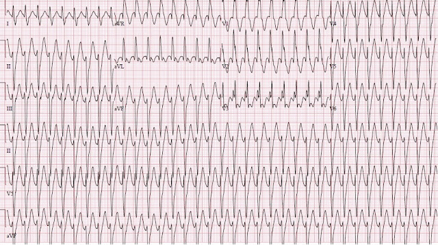
When this didn't help she presented to the ED and had this ECG recorded: 


Here is a higher resolution image, but missing V4-V6:
There is regular monomorphic tachycardia with RR interval of about 0.285 sec (285 ms), for a rate of 210 bpm 
QRS duration is about 120 ms.
What do you think?


Comment: This is a wide complex tachycardia (but only minimally wide!).  

Differential Diagnosis

NOT atrial fibrillation because it is regular.  

NOT polymorphic VT (Torsades de Points, caused by Long QT or Catecholaminergic VT) because all the QRS are identical.

1. SVT with aberrancy
2. AV nodal reciprocating tachycardia (AVRT, antidromic SVT, using an accessory pathway, as in WPW).
3  Ventricular tachycardia (VT)
      a.  "idiopathic" fascicular VT, which has a structurally normal heart
      b.  "Standard" VT, which occurs in a structurally ABnormal hearts such as cardiomyopathy

So let's think it through: 

1. SVT with aberrancy:

A. Sinus tachycardia (a form of SVT) with aberrancy:  Sinus tach can beat this fast in a child.  But there are no P-waves.  

B. Paroxysmal SVT (PSVT) with aberrancy.  

The morphology is similar to RBBB (large R-wave in V1, large S-wave in V6) plus left anterior fascicular block (LAFB) (rS in II, III, aVF, and qR in aVL, which gives it a left axis deviation, away from inferior leads).   

Is it PSVT with RBBB + LAFB?   Unlikely.
       i.    There is absence of rsR' in V1,  typical of RBBB.   
      ii.    QRS duration of 120 ms is atypical for RBBB.  
      iii.   RBBB Aberrancy implies a refractory right bundle.  Although the right bundle is the bundle most likely to be refractory, a child of 12 years should be able to fully conduct at a rate of 210.   

Thus, SVT with aberrancy is unlikely.

2. Antidromic AVRT

Antidromic AVRT travels down through the accessory pathway and up through the AV node.  After traversing the accessory pathway, it must transmit through myocardium (not through specialized conducting Purkinje fibers), which is slow.  Thus, there should be an initial wide depolarization, akin to a delta wave. 

If you thought it was AVRT, it would be easy to test with adenosine.  AVRT depends on the AV node for its circuit and will be terminated by an adequate dose of adenosine.

3.  Standard VT (structurally abnormal heart)

Unlikely in a healthy child who has been playing sports all day.  But it is possible to have myocarditis or other unknown cardiomyopathy.  Standard VT is rarely this narrow, at only 120 ms, though the age of the patient could narrow it.  A bedside ultrasound assessment of would be helpful: normal heart and good contractility would be strong evidence against this diagnosis.

4. "Idiopathic" Fascicular VT (structurally normal heart).  ["Idiopathic" VT is not really idiopathic any more, but rather is known to be due to reentry.  Most are posterior fascicular VT or right ventricular outflow tract VT (RVOT).]

Posterior fascicular VT is a wide complex tachycardia with RBBB and LAFB morphology. It initiates in the posterior fascicle.  A re-entrant rhythm that starts in the posterior fascicle will initiate a rapid depolarization down that fascicle (resulting in a short duration r-wave in inferior leads), then an upward directed depolarization through myocardium (wide) to the anterior-superior LV (resulting in S-waves in inferior leads and an R-wave in aVL).  It will also transmit to the right ventricle and result in an RBBB-like morphology.  

One must also consider RVOT, right ventricular outflow tract tachycardia.  However, the morphology is wrong for RVOT (see below).

A normal bedside ultrasound would be helpful.


Case Progression, as described by the treating physician:

"My initial thought was this was either SVT with aberrancy or V-tach. She was stable with an SBP of 146. Her only complaint was shortness of breath but she was not in any visible distress though she appeared anxious. I asked about family history and she did not have any family history of cardiac problems or arrhythmias. She denied any stimulant use or ingestions." 

"The rate was regular so I decided to try adenosine. I did this first with 6 mg then with 12 mg. Nothing happened, not even a decrease in HR, and I thought that this could be V-tach so I called the pediatric cardiologist at Children's who told me I was pushing the medication wrong and that I needed to do it with a three way stop cock. He said to keep upping the dose until the rhythm broke." 

"I didn't think this sounded right but I did it anyway (in retrospect I should have insisted he look at the EKG first). When this didn't work he finally looked at the strip and agreed that this could be fascicular V-tach. We discussed transfer and he suggested Verapamil. I did give her a 5mg dose and she rapidly converted (2nd EKG). She remained stable and was transferred to Children's. She will have an ablation next month."

"Can you comment and let me know if I should have been immediately onto V-tach?   I don't think Adenosine was the wrong thing to try first. If a patient like this were unstable I would have tried electrical cardioversion. " 

"Any thoughts other than this?" 

"How did you know when I showed it to you that it was from the posterior fascicle?"

Here is the post cardioversion ECG:
Sinus Tach, otherwise normal


Comment:

I knew when I looked at the ECG that it was fascicular VT because of all the features discussed above.  Adenosine certainly will not hurt this, but it will not help either.  Had it been AVRT, adenosine would probably have converted it.

Verapamil in VT: One might be squeamish about giving such a strong negative inotrope as verapamil to someone with VT.  This is due to the fact that most VT occurs in structurally abnormal hearts, especially in ischemic cardiomyopathy, with a low ejection fraction.  Giving verapamil to a patient with VT due to cardiomyopathy could indeed be disastrous.

Thus, our minds associate disaster with verapamil and VT.  In fascicular VT, we should not carry this association.  Of course, if you are hesitant about giving verapamil, just confirm good LV function (good contractility) with bedside ultrasound.

Just for contrast, here is another "idiopathic" VT.  This is a case of right ventricular outflow tract VT (RVOT VT).  RVOT is adenosine responsive.  I presented the case here.
Notice that this tachycardia, in contrast to the above, mostly depolarizes toward the inferior leads.  Late depolarization is also towards the left (wide R-waves in V5, V6), more akin to left bundle branch block. 

Both RVOT and posterior fascicular VT occur in structurally normal hearts and are usually very well tolerated.

Fascicular VT from the posterior fascicle is responsive to verapamil.   RVOT, by contrast, is indeed responsive to adenosine.  Therefore, I would not be surprised it if often gets misdiagnosed as SVT with aberrancy after it converts with adenosine.

Learning Points:

1. Wide complex tachycardia in a child with an otherwise normal heart is likely to be one of the "idiopathic" VTs such as fascicular VT.

2. Fascicular VT (not RVOT) originates in the posterior fascicle, and therefore has RBBB/LAFB morphology.

3. Fascicular VT usually has a relatively narrow QRS (up to 140 ms), whereas VT in a structurally normal heart usually has a wider QRS (not always!).

4. Fascicular VT is verapamil responsive.

5. If you can, ascertain good contractility before giving verapamil.

6. Adenosine will often work for RVOT, which has an LBBB morphology with an inferior axis.


Thursday, July 14, 2016

Dyspnea and Convex ST elevation, Marked LVH, with Bedside Echos

Case 1.

Chief complaint: A 60-something African American male with 5 days of increasing SOB with dyspnea on exertion.

This male in his 60's has a PMH of CAD with MI and CABG, HTN with LVH, hyperlipidemia, and mild HF with only moderately reduced ejection fraction (and some diastolic dysfunction as well).

He presents with 5 days of worsening shortness of breath with orthopnea as well as chest pain.  His BP is 191/90.  He also has a history of venous thromboembolism and has not been taking his anticoagulants.  He was also off of his BP meds (lisinopril, amlodipine and carvedilol).

Here is his ED ECG (ECG #1):
There is 2-3 mm of ST Elevation in V2.  
There is an upwardly convex ST segment in V2 and V3.
The computer read ****STEMI****Of note, the S-wave in V2 is almost 50 mm. This is severe LVH.
Thus, the ST/S ratio is no more than 0.06 (6%)
Note there is also a very prominent negative P-wave in V1, diagnostic of left atrial enlargement and supportive of severe LVH.


Here is his previous ECG (ECG #2):
Here all ST segments are concave upward.  There is slightly less ST elevation.  There is no convexity.
Thus, the convexity is new.
I cannot explain the marked difference in voltage from ECG #1.
They both used 1 millivolt per 10 mm in both limb leads and precordial leads
Bedside ultrasound:

The lung ultrasound (not shown) had B-lines, all but diagnostic of pulmonary edema.

Bedside ultrasound parasternal long axis view is shown below.  


There is severe LVH.  There appears to my eye to be less than optimal systolic function and also less than optimal LV diastolic function.


Here is the short axis view:

Same interpretation as above.


Here is the chest X-ray:


There is obvious pulmonary edema.


Clinical Course:

--The D dimer was barely elevated at 327 ng/mL (upper limit of normal: 230 ng/mL).
--The initial troponin I was 0.083 ng/mL (99% = 0.030); this patient does not have baseline elevated troponin (does not have chronic myocardial injury, which is not uncommon in patients with heart failure), so this was a new troponin elevation.

That is to say, there appears to be acute myocardial injury. 

--The Creatinine was 1.5 mg/dL, with a GFR of 69 ml/min (low).
--The NTproBNP was 1564 pg/mL (slightly elevated -- normal for patients with GFR less than 60 is 1800 pg/mL).

The emergency physicians correctly interpreted the ECG repolarization abnormalities as being entirely due to LVH combined with some injury due to acute heart failure exacerbation and hypertension, and understood that the newly elevated troponin (with rise and fall) was due to demand ischemia (type 2 MI) due to hypertension and acute heart failure.

They administered furosemide.  

Learning point: 
1. ST Elevation with Convexity in the presence of LVH does not necessarily mean STEMI
2. The ST/S ratio in STEMI would be significantly higher.  

See discussion at this post: 

LVH with anterior ST Elevation. When is it anterior STEMI?


Clinical course continued:

They gave furosemide and obtained a CT pulmonary angiogram which ruled out pulmonary embolism.  He was not given any BP management.

Comment on management:

1. With B-lines and this chest x-ray and only a minimally elevated D Dimer, CTPA is unnecessary.  The diagnosis is clearly acute decompensated heart failure.

2. This patient should get some immediate treatment to lower blood pressure (afterload reduction).  IV nitroglycerine is best, but requires ICU monitoring. For a patient who is only moderately ill and does not require an intensive monitoring, a good choice, especially for African Americans with renal dysfunction, is afterload reduction with IV hydralazine (see references below).   Often, in a patient who has not been taking his/her antihypertensive, I simply give those medications immediately in the ED. As this usually includes either an ACE inhibitor or an angiotensin receptor blocker, this is often sufficient.

Vasodilator Therapy of ADHF
http://circ.ahajournals.org/content/123/21/2414
http://www.ncbi.nlm.nih.gov/pmc/articles/PMC1808524/


Clinical Course continued:

Subsequent troponins were 0.078, 0.076, 0.064

A formal echo showed:

Decreased left ventricular systolic performance - mild.
The estimated left ventricular ejection fraction is 45-50%.
Regional wall motion abnormality-anterior and septum.
Regional wall motion abnormality-inferior.
Left atrial enlargement, moderate.
Diastolic septum measurement = 1.25 cm (LVH)
 Based on Doppler indices, the LV filling pressure is markedly elevated.

The patient improved simply by re-starting his hypertension medications, lowering his BP, and with one dose of furosemide.

2.5 weeks later, after recovering from acute decompensated heart failure, his ECG continued to show high voltage and convexity, although he was recovered from the acute heart failure.


Case 2:

A middle-aged patient with a history of severe persistent asthma, heart failure with preserved ejection fraction (HFpEF), and pulmonary hypertension presented presented very short of breath.  Here was her first ECG (ECG #3):
The computer read this as ****STEMI****
Note the marked ST elevation and the convex ST segments.

This is her previous ECG from earlier in the year (ECG #4):
Much less ST elevation and less convexity


A previous formal echo showed:

Hyperdynamic systolic performance.
The estimated left ventricular ejection fraction is 80 %.
There is no left ventricular wall motion abnormality identified.
Left ventricular hypertrophy concentric severe

She improved with therapy for asthma in the ED and had this ECG recorded 5 hours later (ECG #5):
Improved ST Segments and convexity has resolved.


There was no myocardial infarction.  All troponins were negative.  She improved with therapy for asthma and was discharged from the ED.


Learning Points:

1. Patients with LVH who are in acute respiratory distress, especially with elevated BP and/or acute decompensated heart failure, frequently have convex ST elevation.  This does not imply STEMI.

2. The ST/S ratio in STEMI with LVH is not established.  The article by Armstrong et al. which is discussed in the referenced blog post above did not have appropriate methodology and their ratio of 0.25 is far too high.  There certainly should be a ratio higher than 0.10 and probably higher than 0.15.  (Armstrong EJ, Kulkarni AR, Bhave PD, et al. Electrocardiographic Criteria for ST-Elevation Myocardial Infarction in Patients With Left Ventricular Hypertrophy. Am J Cardiol. 2012;110(7):977-983. doi:10.1016/j.amjcard.2012.05.032


Friday, July 8, 2016

A 65 Year Old Man with Chest pain and Precordial ST Elevation

This case was sent by Sam Ghali (@EM_RESUS)


A 65-year-old gentleman presented to the ED complaining of chest pain.  He asked my thoughts on his presenting ECG:
Computerized QTc is 409 ms
What do you think?


Here is my response:

Strongly suspect normal variant or chest leads placed too high.
Possibly STEMI, but I strongly doubt.
In my life, I have seen one saddleback (such as this) that was a STEMI.
Outcome?


Here is Sam's analysis and outcome:

The obvious question at hand here: Is this acute LAD occlusion? 

There is an rSR' in V1 and S-waves in the lateral leads, but QRS less than 120 ms, thus an incomplete RBBB. There is significant ST-elevation in V1-V3, which meets ACC/AHA STEMI Criteria in these leads (greater than or equal to 2.0 mm in an male greater than or equal 40 yo). Also concerning for coronary occlusion, the T-waves are upright, large, and broad in appearance (especially in proportion to the QRS). There is inferior ST-depression in II, III, and AVF which in LAD occlusion is reciprocal to anterior ST-elevation. What speaks against LAD occlusion is the very well-developed R-waves in V3-V4. (As the ECG evolves in LAD occlusion, there is a loss of R-wave height as R-waves transition to Q-waves; however you wouldn't necessarily see this early on). There is concave-up ST-elevation which makes occlusion less likely, but by no means rules it out. What is also unusual in this case is the “saddleback” morphology of the ST-segment elevation.

A prior ECG would be helpful here, but there was none. A bedside echo looking at LAD distribution wall motion might also be helpful but due to logistical reasons this too was not an option. The decision was made to activate the cath lab. 

Outcome

The cath revealed chronic total occlusion of the RCA, with good collateral flow. There were multiple 40-50% lesions throughout the LAD and it’s Diagonals, as well as in the Circ. There was no acute occlusion.   Serial troponins were negative.  Echo did not show LVH.

I shared the case with Steve Smith to gain his expert insight, but here are my comments:

1. STEMI Criteria are imperfect - not nearly sensitive nor specific enough for strict use

2. Even with other ECG features taken into account, diagnosing acute LAD occlusion can be very difficult. The Subtle Anterior STEMI formula which may provide diagnostic guidance here, technically should not be used in the presence of inferior ST-depression.

3. While “saddleback” ST-elevation is less likely to be coronary occlusion, it is still possible for STEMI to have this morphology.

4.  Ultimately, if after all measures there is still concern for acute LAD occlusion, the diagnosis may be best excluded in the cath lab.

Smith comments:

This is saddleback ST elevation, such as one commonly sees in LVH or in type 2 Brugada.  It can also happen if chest leads are placed too high.  It is rarely due to STEMI.  (I was a bit surprised to hear that the echocardiogram did not show any LVH).

The inferior ST depression is probably a false positive.  If you look at the PR segment, it is downsloping.  This is a frequently encountered atrial repolarization wave, and this wave is persistent after the end of the QRS, causing a mimic of ST depression.

Atrial Repolarization wave mimicking ST depression:
You can read about it at this post.
You can watch a K. Wang video about this at this post.

Thus, the formula can be used.  The R-wave amplitude in V4 is the single most powerful predictor of early repol vs. LAD occlusion: a high amplitude, which here is very high at 25 mm, goes strongly against LAD occlusion.  The formula value here, using STE60V3 of 2.5 mm, is very low at 18.97.

Even if the QTc were very long, at 460 ms, the formula value would still be very low at 21.98 (less than 22.0), which would all but rule out LAD occlusion.

That said, one cannot entirely rule out STEMI with the formula and such a case should always of course be approached carefully.

If available, I would do a stat formal echo rather than activate the cath lab.


Friday, July 1, 2016

Middle-aged Diabetic with Syncope, Diaphoresis, Nausea, and Dizziness

This middle-aged male with history of 3 previous stents, 2 in the RCA, had Syncope, Diaphoresis, Nausea, and Dizziness.

He called 911 and the medics recorded an ECG that looks just like his first ED ECG:
There is inferior ST elevation and reciprocal ST depression in I and aVL, with reciprocal T-wave inversion in I and aVL.


This ECG was texted to me (while out on a beautiful point on an island!) with the question: "Would you activate the cath lab?"

I responded: "Yes or at least get stat echo.  It is 90% certain to be occlusion."

Outcome: The coronaries were clean.  The troponins were negative.  The subsequent ECGs did not evolve (this latter is the best evidence that there was no MI, as transient ACS can have all of the following negative: Cath, troponins, and Echo).

It turns out that the patient had a similar false positive activation 1/2 year ago because this is his baseline ECG.  

He must carry a copy with him wherever he goes!!

Sometimes you just have to have a false positive.  You must activate anyway.  You cannot let an ECG like this go without action unless you know it is the baseline.

If there is any doubt, it would be reasonable to find an old EKG and/or to get an emergent echo.

Pretest probability: This also points out (again) that when patients do not have chest pain, especially if they do not at least have dyspnea, the probability that their ECG represents a STEMI decreases considerably.


Recommended Resources