Thursday, December 27, 2012

Before diagnosing STEMI, try to find a previous ECG!

A man in his 50's complained of typical chest pain.  Here is his ED ECG:

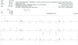
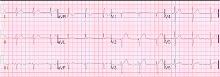
There is a Q-wave in V2, with ST elevation and hyperacute appearing T-waves in V2 and V3.  This is all but diagnostic for anterior STEMI.  The anterior STEMI equation should not, strictly speaking, be used, because the presence of a Q-wave makes STEMI very likely.

The physician was about to activate the cath lab.  However, he found out that the patient had been seen in the ED for chest pain 6 months prior, and had the following ECG:

Nearly identical to the first.  However, the first one has slight straightening of the ST segments in V2 and V3, which are suspicious.

The patient had been ruled out by serial troponins at the previous hospitalization.  He had not had an echocardiogram or any stress testing or angiogram.  The etiology of the abnormal baseline ECG is uncertain.  Is it due to a previous anterior MI?  Very likely.

Because there was no significant change, the cath lab was not activated.  Serial ECGs were recorded and showed no evolution.  The patient ruled out for MI by serial troponins, but, again, did not have a stress test or echocardiogram.

Lesson:

1. Always check old ECGs, if available, when there is a subtle ECG with possible STEMI.
2. Serial ECGs can help to clarify the situation.

Wednesday, December 19, 2012

Middle Aged Male with Chest Pain and Previous MI

A middle aged male with h/o CAD presented with chest pain.  He had a recent previous stent in the circumflex.  Here is the ECG at time = 0:

What do you think?


Here is the previous ECG from one month prior:
Now  what do you think?


The first ECG shows Q-waves in lateral leads, and T-wave inversion as well, wit ST elevation in I and aVL.  There is ST depression in V2-V5, 1 mm in V3, diagnostic of posterior STEMI.  Thus, this is posterolateral STEMI.  The Q-waves may lead one to believe that this is a subacute STEMI with a prolonged duration, long enough to result in infarction in addition to injury.  But the pain duration was only 1 hour.

Comparison with the previous ECG confirms that this is acute STEMI superimposed on a previous lateral MI (old lateral Q-waves, old T-wave inversions).  The ST elevation in I and aVL is definitely new.  And now it is clear that the ST depression in V2-V5 is very marked, as there is 3.5 to 4 mm of relative ST depression in V3.

It is not clear that the treating physicians saw the previous ECG.  They were uncertain of the diagnosis and called  an immediate cardiology consult.  Hydromorphone relieved all the pain, which helped ease the anxiety of the physicians.  The cardiologist arrived  immediately, but did not immediately activate the cath lab.  They ordered this repeat ECG at time = 51 minutes:

Now it is very clear that there is an acute posterior STEMI.  


An unusual feature of this case is the persistent negative T-waves in the affected leads.  Normally, with occlusion of the infarct-related artery, negative T-waves from previous MI will become upright (pseudonormalization).  So one would expect, if this is indeed STEMI, upright T-waves in I, aVL, V5 and V6.

An immediate echocardiogram was ordered which confirmed new posterolateral wall motion abnormality and old anterior and apical wall motion abnormality.  The patient underwent PCI with stent of occluded proximal circumflex near the ostium of the first obtuse marginal where the previous stent had been placed.  The maximum troponin I was 80 ng/ml.

Here is the post-PCI ECG:
Notice the tall and large precordial T-waves.  These are what I call Posterior reperfusion T-waves.  They are the analog of Wellens' T-waves and if recorded from the posterior wall would look like Wellens' T-waves.  But because the leads are over the anterior wall, then they are large and upright, rather than inverted!

Opiates in Acute Coronary Syndrome:

Opiates are associated with 1.5x mortality in ACS.  Why?  Probably because they lead the physician to a false sense of security.  In this case, the absence of any further chest pain was reassuring to the physicians.

See this reference with full text link:


Friday, December 14, 2012

STEMI: Which coronary artery is occluded?

This middle aged male with chest pain had this recorded by the medics at t = 0:
ST elevation in I and aVL and reciprocal ST depression in III, but also with STE in V2, and subtle ST depression in V3 and V4

What artery do you think is occluded?  Answer below.


The medics activated the cath lab prehospital, and then recorded this at t = 8 minutes:
There is now more STE in I and aVL, but also new STE in V3

In the ED, this was recorded at t = 28 minutes:
Now there is STE throughout the precordial leads.


The pattern of ST elevation in I and aVL and V2 (also usually with some ST depression in V3-V5) has been described as a "Midanterolateral MI" (see article below) due to occlusion of the first diagonal (branch of the LAD, also known as D1, or LADD1) artery.  The D1 branches off from the LAD to the lateral wall and may also supply some of the anterior wall.

In this case, as time and ischemia progressed, this became a full anterolateral MI, but the infarct-related artery was indeed a very large LAD D1.  It was rapidly treated and the maximum troponin was low, but there was an anterolateral wall motion abnormality on Echo.

Literature:

Isolated mid-anterior myocardial infarction: a special electrocardiographic sub-type of acute myocardial infarction consisting of ST-elevation in non-consecutive leads and two different morphologic types of ST-depression

Sclarovsky S. International Journal of Cardiology. Volume 46, Issue 1, August 1994, Pages 37–47


Abstract


We describe eight patients with a distinct electrocardiographic pattern of anterior wall myocardial infarction characterized by three main features: (1) a pattern of ‘transmural ischemia’ (ST-elevation with positive T-wave) in nonconsecutive leads: aVL and V2, and two different types of ST-depression; (2) a pattern of ‘true reciprocal changes’ (ST-depression and negative T-wave) in III and aVF; (3) a pattern of ‘sub-endocardial ischemia’ (ST-depression with positive T-wave) in V4–5, while ST in V3 was either isoelectric or depressed. We characterize the electrocardiographic features and correlate them with the echocardiographic, radionuclide, and angiographic data. All patients admitted to the coronary care unit from January 1990 to April 1992 with evolving acute myocardial infarction were evaluated prospectively. Patients whose admission electrocardiogram met the description above were included. The electrocardiographic evolution, echocardiographic, Technetium MIBI tomography, and coronary angiography are described. Of 471 patients with acute anterior wall myocardial infarction, admitted to the coronary care unit during the study period, eight patients met the inclusion criteria (1.7% of acute anterior wall myocardial infarction). Echocardiographyic studies revealed mid-anterior hypokinesis in two patients, anterior and apical hypokinesis in one, and no wall motion abnormality in four patients. Technetium MIBI tomography, done in five patients, was consistent with mid-anterior or mid-anterolateral infarction without involvement of the septum or apex. Coronary angiography, performed in seven patients, demonstrated significant obstruction of the first diagonal branch in all of the patients. In four patients, the diagonal occlusion was the only significant coronary lesion in the left coronary artery. Conclusion: Most of the anterior myocardial infarctions also involve the septal and apical regions. Anterior wall myocardial infarctions limited to the mid-anterior or mid-anterolateral wall, without apical or septal wall involvement are relatively rare. This study describes a special electrocardiographic form of anterior wall acute myocardial infarction. This distinct electrocardiographic pattern represents true mid-anterior wall myocardial infarction, caused by occlusion of a first diagonal branch of the left anterior descending coronary artery. The septal and apical regions are not involved because the blood supply via the left anterior descending artery is not interrupted.


Friday, December 7, 2012

Two patients with chest pain and ST elevation. Are they STEMI?

Both of these patients presented with chest pain.  What do you think of the ECGs?


For the answer, go to the free pdf of this paper I authored in Annals of Emergency Medicine, p. 52 (the 8th page of the paper).   There is a full explanation of the formula which helps to differentiate these two entities. 

There is an online excel spreadsheet on the right sidebar of the front page of this blog which can be used to calculate the formula value and help you to assess your patient's ECG.

The picture below shows where to find the online calculator.  It is important to read the limitations in red above the calculator:


Monday, December 3, 2012

Back pain radiating to the chest in a man in his 40's

A man in his 40's presented with severe back pain radiating to the chest.  Here was his ECG, with pain:

What is your diagnosis?


This is diagnostic of hyperacute T-waves and LAD occlusion or subtotal occlusion.  There are subtle de Winter's T-waves in V4-V6, in which the large T-wave is preceded by ST depression.

Apparently, it was not recognized (which is not unusual, this is subtle and difficult).  Aortic dissection was on the differential diagnosis, and a CT of the chest ruled this out.  Troponins were negative, the pain was resolved, and the patient was discharged.

He returned the next day with 9/10 back pain.  Here was his ECG:

What do you think?


The fact that the T-waves are so different from the previous day's confirms the ischemia of the previous day.  There is ST segment elevation in precordial leads.  Is it normal variant ST elevation ("early repolarization")? 

Even without a previous ECG for comparison, this is unlikely to be normal STE because of the poor R-wave progression.  In our study of early repol vs. subtle anterior STEMI, only 5% of early repol had an average R-wave amplitude from V2-V4 of less than 5 mm.  Here it is 1 + 2 + 9 = 12 divided by 3 = 4mm average.   If you use the formula (see sidebar) which incorporates the ST elevation at 60 ms after the J-point in lead V3, and the QTc (393ms), then the result is 23.8, which is greater than 23.4 which indicates anterior STEMI.

His pain resolved spontaneously, and he had the following ECG:

This is now obviously diagnostic of MI, and with T-wave inversion and absence of pain, the artery is likely to be open.  The Q-waves indicate that the MI has probably been going on for quite some time.


The troponin returned positive this time and the patient went for cath and had subtotal stenosis of the LAD just distal to the first diagonal.  It was stented.

I don't know the peak troponin, or echo results, or how much myocardium was lost.  But here again is a "NonSTEMI" that really should be treated like a STEMI.  The ECG 1 week later is consistent with significant myocardial loss:

Near total loss of R-waves from V2-V4

Monday, November 26, 2012

Full Text of Left Bundle Branch Block Article Now Free Online

Smith SW et al.  Diagnosis of ST-Elevation Myocardial Infarction in the Presence of Left Bundle Branch Block With the ST-Elevation to S-Wave Ratio in a Modified Sgarbossa Rule

There is one rather glaring, but inconsequential, error in the article.  I'm interested to see if you pick up on it, so I won't say what it is yet. 

And, of course, we will publish an erratum later, unless there is a letter to the editor to which we will respond.


Don't forget:  Dr. Smith's ECG Blog will be live online at 1:30 PM Central Time on Thursday, Nov. 29, 2012.  Video will be broadcast directly on the blog site, and you may call in to ask a question using Skype.  The Skype ID is: smithecg 

Wednesday, November 21, 2012

An Emergency Physician Texted Me This ECG, Asking for Help in Interpretation and for Advice in Management

I  was in a meeting and received a text message: "can you look at an ECG?"

I texted back to take a photo and send it.

The patient is a male over 40 years of age with central chest discomfort for several  hours.  Here is the ECG, as texted:

What is your diagnosis?


Answer

I called her back and this is what I said:


“This is a definite posterolateral MI, with an occluded artery, probably an OM, but it is not technically a STEMI because there is not 1 mm in 2 consecutive leads.  He needs to go to the cath lab ASAP.”


How did I know this?

Because there is BOTH: minimal STE in I and aVL with reciprocal ST depression in III, AND ST depression in V2 and V3. 

Combine this with sudden onset of substernal chest pain and you have  a certain posterolateral MI.

She called the cardiologist on call and emphasized the need to go ASAP to the cath lab.  The patient's pain resolved completely with intensive medical therapy, and so the urgency was less.  Within a very short time, the patient underwent an angiogram that showed a 99% OM-1 occlusion and and 85% RCA.  Both appeared "hazy"; both appeared to be culprits and both underwent stenting.

A subsequent echo was normal, without wall motion abnormality.  Remember this! 

The troponin I peaked at 58 ng/ml, so this was not a small MI.


Learning points:

  • 25-30% of "NonSTEMIs" have an occluded infarct-related artery at 24 hours after presentation.  (1, 2, 3)
  • Many more are occluded at the time of presentation.
  • This ECG would be read as "non-diagnostic" by the vast majority of interpreters.  There are many "nondiagnostic" ECGs that really are diagnostic if scrutinized closely.
  • Cardiac ultrasound done after resolution of ischemia is only moderately sensitive for MI.


References

1.  Wang T, Zhang M, Fu Y, et al. Incidence, distribution, and prognostic impact of occluded culprit arteries among patients with non–ST-elevation acute coronary syndromes undergoing diagnostic angiography Am Heart J 2009;157(4):716-23.
2. From AM, Best PJM, Lennon RJ, Rihal CS, Prasad A. Acute Myocardial Infarction Due to Left Circumflex Artery Occlusion and Significance of ST-Segment Elevation. Amercan Journal of Cardiology 2010;106(8):1081-5.
3.  Pride YB, et al.  Angiographic and clinical outcomes among patients with ACS presenting with isolated ST-Segmment Depression: A TRITON-TIMI 38 Substudy.  Journal of the American College of Cardiology: Cardiovascular Interventions 2010;3(8):806-11.


Friday, November 16, 2012

Chest Pain and Right Bundle Branch Block. What is the Diagnosis?

A male in his 70's with no h/o MI or CAD presented with new onset chest pain.  Here is his ED ECG:


There is sinus rhythm and right bundle branch block, and ST elevation in V1-V3. 
Normally, RBBB has ST depression in right precordial leads, especially V2 and V3 (see ECG below).  Sometimes it is isoelectric.  But if there is any ST segment elevation, as in this case, it is STEMI (or RBBB with old MI and persistent ST elevation) until proven otherwise.

Baseline RBBB (normal RBBB, no STEMI). Note that there is a large R' wave in V1-V3 and discordant (opposite direction of QRS) ST segments. (Thanks to K. Wang's EKG Atlas for this image)

The emergency physician was very worried about STEMI, called the cardiologist on call, and they performed an immediate bedside echo which confirmed absence of anterior wall motion.  Immediate angiogram was done and confirmed LAD occlusion, which was opened and stented.

Here is another example.

Here is a 3rd example.

Here is a 4th example.

Friday, November 9, 2012

Wide Complex Tachycardia. What is the Diagnosis?

A man in his 40's with a h/o coronary disease complained of sudden dizziness and chest pain.  Medics found him with a high pulse, and rhythm strip and prehospital 12-lead (not shown) had a wide complex at a consistent rate of 135.

On arrival, here is his initial ED ECG, and is identical to the prehospital ECG and at the same rate:

There is a regular, wide complex.  No p-waves are seeen.  The wide complex has a right bundle branch and left anterior fascicular block morphology.  No flutter waves are apparent.


So this is most likely SVT (PSVT) with aberrancy (RBBB) because: a) the typical RBBB pattern makes VT unlikely b) the absence of p-waves and the constancy of the rhythm makes sinus tach unlikely and c) flutter should have something that could be construed as flutter waves in leads II and/or V1.

Alternatively, it could be posterior fascicular ventricular tachycardia.

Therefore, adenosine was given: the patient responded with several seconds of asystole.  No underlying flutter waves were uncovered and no p-waves either.  The rhythm gradually returned to its baseline at a rate of 135.  So it is not VT, but it was still puzzling.  Either the PSVT was broken and restarted, or there is sinus tachycardia.

Having remembered Christopher Watford's post on EMCrit about the Lewis lead, I Googled "Christopher Watford" and found the post.

Just as it describes, I then:

  1. Placed the Right Arm electrode on the patient’s manubrium.
  2. Placed the Left Arm electrode on the 5th intercostal space, right sternal border.
  3. Placed the Left Leg electrode on the right lower costal margin.
  4. Monitored Lead I.
This is the rhythm strip that resulted:
Now p-waves are obvious (and large!)

 By changing the lead orientation, one can detect atrial activity with much higher sensitivity.  Our electrophysiologist learned of this age-old technique (first described by Sir Thomas Lewis in 1931) technique from his mentor, and also uses it in wide complex tachycardia to detect atrioventricular dissociation (when you see AV dissociation in wide complex tachycardia, it is diagnostic of ventricular tachycardia).

The link to Christopher's interview provides all the resources you need to learn more about the Lewis Lead.

Some Final Comments:

I have added this because of some comments/questions of whether I might treat with verapamil because this looks like verapamil sensitive idiopathic VT:

While this does look like verapamil sensitive idiopathic VT, it does have a typical bifascicular block pattern.  The only thing atypical about it is the T-waves in V2 and V3, but the QRS pattern is typical for RBBB + LAFB.

The patient's baseline ECG looks exactly the same, except that it is slower (93) and the p-waves are obvious.  The Lewis leads proved that it was indeed sinus tach.  And with time and fluids and anxiolytics, the rate did trend down.

Sinus tach is usually compensatory to some underlying illness.  Maybe the patient has dehydration, sepsis, hemorrhage, or PE.  If you were to give verapamil to someone with sinus tach secondary to underlying pathology, you would harm them and perhaps provoke hypotension and shock.

Thus, I would be very careful about giving verapamil to someone with such an ECG until you have proven that it is not sinus tach.

In fact, this is what we did.  My resident wanted to treat the presumed SVT with verapamil because it had apparently recurred after adenosine.  I expressed my concerns as above, and that is when we did the Lewis leads.

Outcome:

The etiology of the patient's sinus tachycardia was not discovered.  He later returned to the hospital for recurrent bouts of idiopathic sinus tach.

Here is his ECG 24 hours later, at a rate of 118:

P-waves at this slower rate are now clearly visible.
The QRS morphology is identical, confirming that this rhythm had to be supraventricular.
The supraventricular rhythm was sinus


Here is an ECG from 2 weeks later:
Again, this is clearly sinus rhythm with RBBB and LAFB.


Learning Point:

When SVT with aberrancy or VT are suspected, both may be ruled out by diagnosis of sinus tachycardia using Lewis Leads.  These are simple and fast.

Recommended Resources