Tuesday, December 27, 2011

Subtle Lateral ST elevation. False positive. This diagnosis is hard.

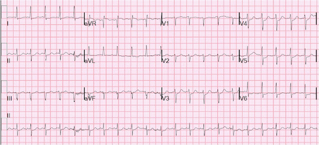
Case 1.  A middle-aged woman presented with severe substernal crushing chest pain radiating to the left shoulder.  This was her presenting ECG:
There is a Q-wave in aVL and Minimal ST elevation in I and aVL (less than 0.5 mm) in the context of a 10 mm QRS.  There is minimal reciprocal ST depression in III.  There are some features of left anterior fascicular block, but  there is not enough left axis deviation to meet criteria for this.  This could be high lateral MI with circumflex or diagonal occlusion.
Management options are to get an immediate echocardiogram, look for old EKGs (there were none), do serial ECGs, or just do an immediate angiogram.  The interventionalist was consulted and he opted to go for immediate cath.  It was negative.

Below are 3 typical cases (cases 2-4) of lateral ST elevation with inferior reciprocal ST depression that were actually circumflex or diagonal occlusions.  In these cases, the findings are not as subtle as in case 1.  But when you look at the last case (case 5), you'll see how subtle real occlusion can be.  

It is important to remember that only about 50% of lateral MI due to coronary occlusion have significant ST elevation, and for this reason the lateral wall is often called "electrocardiographically silent."  Often this is due to low QRS voltage in lateral leads, and because ST elevation is always proportional to the QRS, the ST eleavtion is low voltage.  (See Schmitt et al. Chest 2001;120(5):1540-6, free full text)

Case 2.
Acute Circumflex occlusion (also old inferior MI with Q-waves, and early repol giving anterior STE)

Case 3.
Lateral STEMI with very low voltage QRS in aVL, therefore very low voltage STE in aVL, but best seen by marked ST depression in inferior leads.  Diagonal occlusion.  Remember ST depression in inferior leads is not inferior ischemia, but rather reciprocal to lateral STEMI
Case 4.
Circumflex occlusion with hyperacute T wave in aVL and reciprocal inferior ST depression.  Notice the precordial ST depression of concomitant posterior STEMI.


Case 5a.
D2 occlusion, lateral MI


Case 5b.
After nitroglycerine, inferior ST depression and STE in aVL resolve.  D2 is open by the time of angiography.

Here is one of the most popular posts of all time, also relevant to this:  ST depression does not localize: 2 cases of "inferior" ST depression of high lateral STEMI. 

Monday, December 19, 2011

Ventricular fibrillation on a 12-lead ECG

A 54 year old male suddenly collapsed.  He had not complained of symptoms prior to this.  He received bystander CPR, then was defibrillated when the medics arrived.  He arrived in the ED awake. The following ECG was recorded:
There is sinus tachycardia with diffuse ST depression (I, III, III, aVF, V2-V6), with obligatory ST elevation in aVR (ST depression maximal in leads II and V4 establishes the ST vector as upward and rightward; there must be ST elevation in aVR).

This is typical of NonSTEMI with ischemia from acute 3-vessel ischemia or left main stenosis.  It establishes ongoing ischemia, though sometimes may be residual after cardiac arrest.  So a repeat ECG should be done a short time later in order to establish whether there is, indeed, ongoing ischemia.  Thrombolytics are never given for such an ECG, but angiogram and PCI are indicated if medical therapy alone does not control the ischemia.

Amiodarone 150 mg was given, along with aspirin, heparin, and eptifibatide.  Because of tachycardia and thus a risk for cardiogenic shock, no beta blocker was given.  BP was adequate and so nitroglycerine drip was started.  Clopidogrel (or any thienopyridine) was not used because the ST elevation in aVR makes the probability of CABG high.

15 minutes later, the patient was awake enough to take a history.  He stated that he had ongoing chest pressure, strongly suggesting ongoing ischemia.  The leads were placed for a repeat 12-lead ECG, and we were talking to the patient when the ECG tech said, "Hey, guys, uh......"  We looked up and this is what we saw:

12-lead ventricular fibrillation.  Not seen very often!!  Notice how similar ventricular fibrillation is to torsadeBy the way, the computer read: sinus tachycardia with frequent multiform PVCs!!!!


The patient was awake but becoming obtunded when he was defibrillated successfully with one shock; he did not remember it.

Recurrent v fib in the setting of ischemia is diagnostic of ongoing and uncontrolled ischemia.  The only way to control this is with PCI, if angiogram shows a lesion amenable to therapy.

A second 150 mg dose of amiodarone was given.

Angiogram showed 3-vessel disease and 3 lesions in the LAD which were stented.  He did well.

Saturday, December 17, 2011

Right Ventricular MI seen on ECG helps Angiographer to find Culprit Lesion

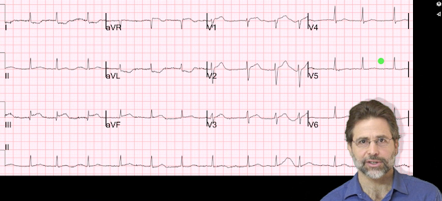
This is a video I made a while back.  I thought it was worth a re-post.  For those who don't have time to watch a video, you'll have to read the ECG as shown on this still frame because I lost it and cannot post it.

See down below for explanation if you don't want to watch the video.

There is inferior STEMI.  But there is also ST elevation in leads V1 and V2.  When you see this, think right ventricular (RV) MI.  The hypotension is further evidence for RV MI.  There was no right sided ECG.

I heard about the case, and saw the ECG, shortly after the patient left for the cath lab.  I called the interventionalist while the patient was on the table and he told me that the occlusion was not in the proximal RCA, but further down.  I asked if he was sure about this, because the ECG would indicate a proximal RCA occlusion with RV MI.  He took another look and realized that the culprit was indeed in the proximal RCA and that the thrombus had embolized distally.  And so he put the stent in the proximal RCA.

Learning point: Even when you have an angiogram, the ECG findings make a difference.


Tuesday, December 13, 2011

This ECG is nearly pathognomonic. What is it?

A 45 yo male with a known history of MI presents with a few hours of chest burning, resolved now.  Here is his presenting ECG:
There is ST elevation in precordial leads.  What is the diagnosis?  See below.  The first troponin I returned positive at 0.467 ng/ml.


This is classic Left Ventricular Aneurysm morphology, otherwise known as persistent ST elevation after old MI.  There are QS-waves in V2-V4 (QS-wave is a single negative deflection without any R-wave), moderate ST elevation, T-waves are not tall and may be (as in this case) slightly negative.

"LV aneurysm" morphology is so-called because it is associated with an anatomic aneurysm about 80% of the time.  It is quite common after a completed MI (formerly known as transmural MI), one in which the artery did not reperfuse, nor was the affected wall reperfused by collaterals, so the entire wall is infarcted, throught the full thinkness.    Over time, the scar thins out and bulges outward in diastole (diastolic dyskinesis on echo).  Before the reperfusion era, they were the most common reason for ST elevation STEMI mimic, but now they are much less common than before.

LV Aneurysm can be inferior, anterior, or posterior.  I have never seen a lateral LVA, but I suppose they could exist.  Inferior aneurysm looks very much like acute MI because it does not get QS-waves, but rather QR-waves, which can also be present in acute MI.  I will post a case of inferior aneurysm soon, but here is one.

The chest pain with troponin elevation establishes this as a Non-STEMI.  There is no need to activate the cath lab emergently.  In his case, next day echo confirmed apical and anterior dyskinesis, and he underwent risk stratification with a nuclear stress test, which was normal, and he did not undergo cath.  [This is perfectly appropriate conservative care for NSTEMI if the patient is discharged on maximal medical therapy (statins, beta blockers, aspirin, clopidogrel)].

Here is an old post from 2009 that describes LV aneurysm in detail, and describes an ECG rule I developed to help differentiate it from STEMI.

Here are all the cases I have posted on LV aneurysm.

Friday, December 9, 2011

Subtle Inferoposterolateral OMI, sent home!!

This ECG comes from Tom Bouthillet, who is devoted to good STEMI care, runs the EMS12lead ECG site, and who has also produced an outstanding iPhone/iPad/Android 12-lead ECG challenge App for learning to recognize subtle STEMI and to differentiate STEMIs and look alikes.

Case 1. This was a 70 year old woman who had chest pain while exercising the day before, then developed chest pain on the day of the ECG and called 911.  Here was her prehospital ECG.
There is very subtle ST elevation in inferior leads, with hyperacute T-waves, with subtle reciprocal ST depression in I and aVL, and T-wave inversion in aVL.  There is minimal ST depression in V2.  T-waves in V5 and V6 are perhaps a bit hyperacute, too.
This is diagnostic of inferoposterior OMI

For contrast, let's look at limb leads and V4-V6 side by side with a normal ECG:
Here the contrast in T-wave size is obvious. Normal has a wide range, and not all normal inferior T-waves are this small.  But the ones on the left are clearly too large.
Here the contrast in T-wave size is obvious. Normal has a wide range, and not all normal V4-V6 T-waves are this small.  But the ones on the left appear too large.

The medics did not see this.  The computer did not see this.  The patient was brought to the ED and discharged after ED evaluation.  Unfortunately, we do not have any ED data on this case.  What did the ED ECG look like?  Was there an old one for comparison?  It is hard to imagine that this was a baseline ECG.

I suspect this was a missed OMI.  Most patients with missed OMI at least get admitted to the hospital for a rule out.

Case 2. Here is a similar case in which all the data is available:


A 65 year old woman with no previous cardiac history presented with 2 hours of typical chest pain.

There are inferior hyperacute T-waves, some minimal ST depression in V2, and lateral hyperacute T-waves.  There is 0.5 mm of ST depression in aVL. 
This is diagnostic of inferoposterolateral OMI.


Here is the patient's previous ECG:
It is not entirely normal, but there are no large T-waves anywhere.  This establishes that the presentation ECGs findings are new.

These findings were also not recognized.  The patient was admitted to the CCU.  Troponin I peaked at 63 ng/ml 14 hours later.  Angiography showed an occluded dominant left circumflex.  Echo showed an infero-postero-lateral wall motion abnormality and EF of 55%.

Learning points: Scrutinize the ECG for T-wave size and morphology, especially in reciprocal leads III and aVL.

Sunday, December 4, 2011

Several Cases of ST Elevation from Early Repolarization

As I have pointed out in other posts, I have developed an equation to help in the electrocardiographic differentiation of anterior early repolarization from anterior STEMI.  If the equation [(1.196 x ST Elevation in mm at 60 ms after the J-point in V3) + (0.059 x computerized QTc) - (0.326 x R-wave Amplitude in mm in V4)] has a value greater than 23.4, vs. less than 23.4, it is quite sensitive and specific for LAD occlusion.

Remember: when you are uncertain, look for old ECGs, do serial ECGs, then, if you still need to, you can get an immediate echocardiogram, and if you ultimately cannot be certain that it is not STEMI, then you may have to risk a false positive cath lab activation. That happens.

Case 1.  One of my partners phoned me when I was out.  He was worried about this ECG.  He used his iPhone to photograph it, then sent it to me by text message:


Computerized QTc was 391 ms, STE at 60 ms after the J-point is 2 mm, R-wave is 11.5. I looked at it at said I do not think it is STEMI.  If you apply the equation, the value is 21.7 (less than 23.4, so it is early repolarization).
 I told him that it is very unlikely to represent STEMI.

Here is a previous ECG from one year ago:
The new one looks different from this one, especially in V3, but this can happen in early repol
 This was recorded the next AM, after the patient had ruled out with serial troponins:

Here are all 3 side by side:

Case 2.
A Hennepin residency graduate called me to ask about an ECG.  I happened to be in the ED so I asked her to fax it.  She was worried about diffuse ST elevation and whether there was MI or pericarditis.

There is 4 mm of STE at 60 ms after the J-point (2.5 mm at the J-point) in V2, so it looks scary.  There is 1 mm STE in 2 consecutive of inferior and lateral leads.  So it meets "criteria" for fibrinolytic therapy in every coronary distribution.  But the computerized QTc is 370 ms, the STE at 60 ms after the J-point in V3 is only 2mm, and the R-wave in V4 is 19 mm.  Equation value is 21.08, so this is unlikely to be an anterior STEMI.  Is it lateral or inferior STEMI?  The pronounced J-waves make early repol in inferior or lateral walls much more likely than STEMI.  The absence of reciprocal ST depression in aVL makess inferior MI extremely unlikely.
 As for pericarditis: the ratio of ST elevation to T-wave in V6 is less than 25 percent, so pericarditis is unlikely.  Furthermore, there is no significant PR segment depression.

Case 3.  A 19 yo with stab wound to the chest.  After viewing the ECG, there was concern for LAD laceration.

Again, there is scary ST elevation.  In fact, the ST segments in V2 and V3 are straight, not concave.  In my study, I excluded ECGs with non-concave (straight or convex) ST segments because they are so specific for STEMI.  Nevertheless, if you apply the equation, the value is only 20.52.   The patient ruled out.  The heart was not affected by the stab wound.


Recommended Resources