Friday, December 31, 2021

Should we activate the lab? A simple but important lesson

Written by Pendell Meyers


A man in his 60s called EMS for sudden chest pain and shortness of breath. He was found in moderate respiratory distress, hypertensive, diaphoretic, and hypoxemic. He was given aspirin, nitroglycerin, and placed on noninvasive positive pressure ventilation during transport. Medics recorded a 12-lead and transmitted it to the provider, asking if they wanted to activate the cath lab.

Here is the ECG:

What do you think?


Sinus rhythm. In this EMS ECG, as is true for many EMS ECGs, the machine cuts off the S wave voltage at 10 mm. You can see this visually by the subtle but noticeable squared-off waveform of the S wave in leads V2-V3.

In lead aVL, you can see that the machine does not limit the R waves to 10 mm. So as far as I can tell, this machine only limits negative QRS voltage to 10 mm. 

In limiting the S wave to 10 mm, this feature of the EMS 12-lead ECG significantly changes the visual proportionality of the ST segments and T waves in the affected leads. If this QRS were only 10 mm or less in amplitude, then these STEs and relatively large T waves in leads V1-V4 could be worrisome for anterior OMI.

But after falling for this trick once, you probably will not make this mistake again. You will see easily that this QRS shows significant voltage and morphology of LVH.


So the provider said no, no prehospital cath lab activation for now and we will evaluate the patient immediately on arrival.


The patient arrived and was already much improved from the paramedics' treatments thus far. 


Here was his ED ECG on arrival:


Seeing the full QRS makes all the difference here. With the full QRS complexes available, you can see that it is diagnostic of LVH, with very reasonable and proportionally normal STE and upright T waves in V1-V4, with appropriate T wave inversion and STD in V6, I, and aVL. Many would call this "LVH with strain pattern." If the QRS were normal, this pattern of STE and large T waves in V1, with STD and T wave inversion in V6, could have been indicative of LAD occlusion. 


There was a baseline ECG available on file from many years ago:

I understand that there are differences between the baseline and the current ECG. The T waves are slightly taller in proportion to their QRS complexes. The T waves in V1 and V6 are flipped compared to prior (there is not much LVH "strain" on this older ECG). But my opinion is that they are both versions of LVH without clear signs of OMI, and the change between them is also not specific for OMI. Serial ECGs would be able to prove this theory right or wrong.


The cath lab was not activated.

The clinical impression was acute hypertensive pulmonary edema. (it is somewhat unusual in my experience that such a patient would not be tachycardic on arrival, but who knows, maybe they were on a beta blocker).

He was treated for this and dramatically improved within the hour. 

His initial troponin T was less than 0.01 ng/mL (undetectable, normal less than 0.01 ng/mL), and his serial troponins over the next 12 hours peaked at 0.03 ng/mL, then fell. This would be an extremely low peak level of troponin even with a brief LAD OMI.


Angiogram was not performed during this visit. 

Serial ECGs were reportedly performed, but not available. The progress notes described the ECGs as remaining unchanged. (this is important, because if the patient had suffered transient subtle anterior OMI, then we would likely have seen anterior reperfusion changes including deepening terminal T wave inversions).

He improved and was discharged home.


It is extremely unlikely in my opinion that his presentation represented OMI (but theoretically still possible and not ruled out by the information above). 


Learning Points:

ST segments and T waves can ONLY, EVER be properly interpreted in the context of the QRS that made them. Everything on the ECG is always proportional. Evaluation of raw ST or T wave voltage without consideration of the QRS is useless.

EMS 12-lead ECGs often have a voltage cutoff that may affect visualization of the QRS. This becomes especially important and tricky for patients with LVH, LBBB, etc., who may suffer a false positive cath lab activation due to this effect.

Expert electrocardiographers understand that there can be some differences from a baseline ECG that do not have acutely important meaning. The idea that "baseline ECGs" should remain perfectly static over time is a beginner's concept that the intermediate learner quickly understands to be an oversimplification. In actuality, patients have some fluctuation in their ECG even under normal conditions, change with time, and exaggeration by many clinical features such as tachycardia, hypertension, etc.

Retrospective adjudication of the presence or absence of OMI can be tricky and requires understanding many aspects of the clinical presentation, serial ECGs, troponins, cath findings, etc. This is especially true when no cath was done, however even a "negative" cath doesn't rule out OMI. The case above is one in which many ancillary data points make me quite sure there was not OMI. Most importantly, OMI is NOT defined or adjudicated solely based on an ECG finding - that would be a ridiculous way to classify MIs - and that's why the name OMI doesn't say anything about the ECG! Its about the patient's actual pathophysiology that we are trying to identify and treat!!!

CJEM Open Access OMI Quality Improvement Publication

Just published in the CJEM:


From STEMI to occlusion MI: paradigm shift and ED quality improvement


Jesse T. T. McLaren
H. Pendell Meyers
Stephen W. Smith
Lucas B. Chartier


Wednesday, December 29, 2021

Hyperacute T-waves -- missed. Myocardial Infarction with Non-Obstructive Coronary Arteries (MINOCA) may be due to transient thrombotic Occlusion MI.

Coronary thrombosis (twice in the same patient!!) without a stenosis or even a culprit

Do not miss the last image at the bottom that shows the series of T-waves in V4-V6

I recently had a discussion with an incredibly smart and fantastic ECG and Cardiology expert.  He was skeptical that you can have OMI with Wellens waves without having a major stenosis on angiogram.

I told him I've seen it on occasion and that this happens due to thrombosis of non-obstructive lesions that lead to complete occlusion but that then completely lyse and do not show stenosis by the time of the angiogram.  I said that even if there is not a stenosis, they often show a culprit (ulcerated plaque), but not always.  

The day after that discussion, this case came in: Transient Occlusion MI (Transient OMI) that occurred twice, but without any stenosis or even a culprit.

The case demonstrates how carefully you must read the ECG, and how carefully you must compare T-waves size.  It demonstrates that you must look not only at the angiogram, but to the symptoms, ECG, troponins, and echo for the diagnosis.

You will note that the angiogram on the first presentation manifested an initially unseen occlusive culprit, which was only later seen on angiogram over one year later.

Case

A 40-something woman with DM presented with substernal chest pain that felt like her previous myocardial infarction, 1.5 years earlier.  That previous acute MI was diagnosed as MINOCA (Myocardial Infarction with Nonobstructive Coronary Arteries.)

We will give some details on that previous infarction below.


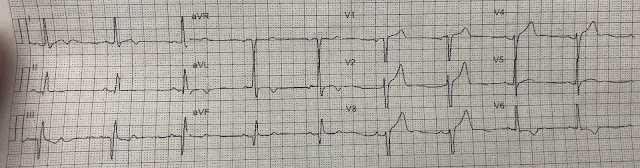
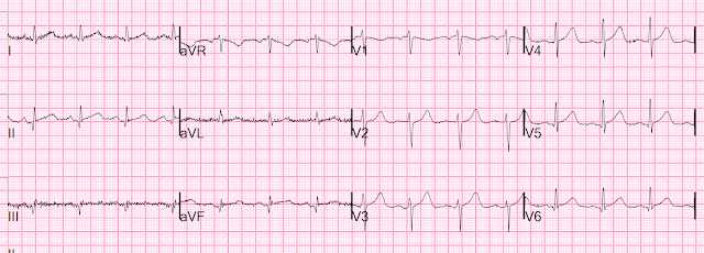
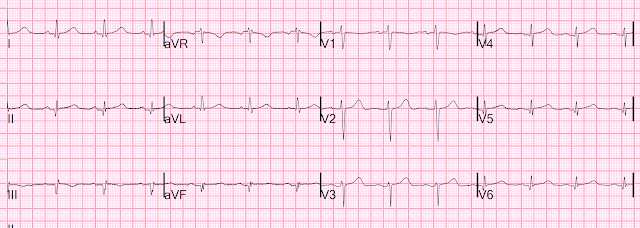
Here is her initial ED ECG (ECG 1):

What do you think?


Smith interpretation: the T-waves in V4-V6 are hyperacute.  These are NOT normal T-waves.  They are large and fat.  The inferior leads look very suspicious as well, with Q-waves and a straight ST segment in leads II and aVF.  This is very suspicious as well.

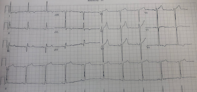
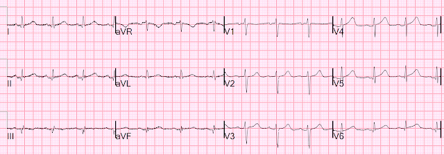
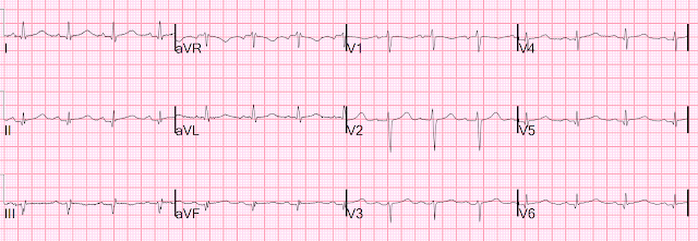
Let's look at her last ECG, just before discharge after hospitalization for the previous MI:
This is completely typical evolution of an inferior Occlusion MI (OMI).  There are inferior Q-waves, slight ST elevation ("coving" or Pardee T waves) with T-wave inversion.  There are also inverted T-waves in the lateral leads V4-V6.  So this previous OMI was inferolateral (I will show the presentation ECG below)

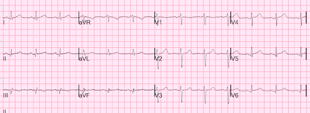
So today's ECG has new upright T-waves in inferior and lateral leads.  Is this pseudo-normalization?  

No!!  The natural evolution of inverted T-waves after acute MI is to become upright over weeks to months.  Even without a new MI, one would expect the T-wave in II, III, aVF and V4-V6 to be upright (but not hyperacute, as here!)  

True pseudonormalization happens within days and is due to acute re-occlusion of a previous occluded and reperfused coronary.
__________
See these 3 cases for LAD pseudonormalization:
__________

The only way you can tell that ECG 1 above has ischemia is to recognize the hyperacute T-waves.  The old ECG only helps you by showing that the Q-waves are old.


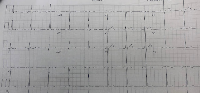
For the prior OMI, here is the presentation ECG:
Inferior, posterior, lateral Occlusion MI that also meets STEMI criteria in inferior leads (STEMI)

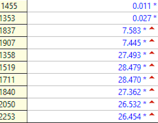
Troponin I profile (contemporary, not high sensitivity) on that previous visit, in ng/mL:
A large acute Occlusion MI

The angiogram at that time showed:

1. Left main: no significant stenosis.

2. LAD: type II vessel that just reaches around the apex. It supplies two diagonal branches. There are luminal irregularities in the LAD but no significant stenosis. Likewise, there is moderate up to 50% disease in D1.

3. LCX: non-dominant. It supplies several tiny OMs and then a large OM.  There are no significant stenoses.

4. RCA: It supplies an RPDA and a small RPLA. There are no obvious stenoses or vessel occlusions.

In other words: Non-Obstructed Coronaries


Formal echocardiogram:

Regional wall motion abnormality-lateral and inferolateral


So this previous visit was MINOCA.


Let's get back to the 2nd presentation.  Again, ECG 1:


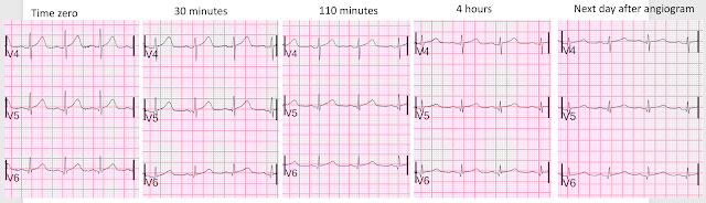
Her pain began to wane and then resolve, and more ECGs were recorded:

Time 30 minutes:
Lateral T-waves are getting smaller in V4-v6

Time 110 minutes:
V4-V6 T-waves smaller still
Inferior T-waves are inverting ("inferior Wellens' waves, or reperfusion T-waves)


Time 4 hours
Now there is complete resolution of the hyperacute T-waves and inversion of inferior T-waves.

However, no one noticed that these T-waves are ischemic, as far as I can tell.  In fact, later interpretations were "She has no current of injury on her EKGs."

The first high sensitivity troponin I was 185 ng/L (this number has a 70% positive predictive value for type 1 MI in our department, but not necessarily for Occlusion MI).

Subsequent trops went to 860, then 2401, then 6658 ng/L.

Since the pain was resolved, and the ischemia was never recognized on the ECG, she was put on heparin and aspirin and angiography was delayed until the next day.

Angiographic findings:


1. Left main: No stenosis.

2. LAD: tortuous. Moderate diffuse disease distally toward the apex. No

obvious focal stenosis. Supplies a diagonal branch without stenosis.

3. LCX: non-dominant. Supplies very small OMs and a medium sized tortuous

LPLA with a very distal 80% stenosis in a small caliber branch that, upon

review of her previous angiogram, had been occluded and is now open.

4. Ramus intermedius: no significant stenosis.

5. RCA: dominant. Supplies an RPDA and small RPLA. No stenosis.

In other words: Non-Obstructed Coronaries


To me, this implies that the PREVIOUS diagnosis of MINOCA was incorrect, as the angiographer now sees an artery that was closed at the previous angiogram but is open now.

But this 2nd angiogram does not show a culprit for the 2nd visit, as far as I can tell.

It is unclear to me if this same vessel is the culprit again.

The hs troponin peaked at 14,143 ng/L.  This is very high and is a typical level for an Occlusion MI [including STEMI (+) OMI].  

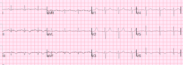
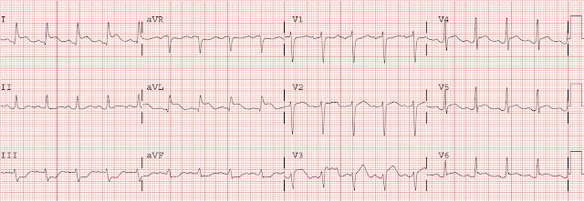
Here is the post angiogram EKG:
All hyperacute T-waves are now normal


Here I put V4-V6 all on one image for the 5 ECGs
Now that hyperacute T-waves in V4-V6 become obvious.  The ones on the far right are normal, after reperfusion.


There was no lesion that could be stented.  She was treated medically for her presumed ACS MINOCA.


MINOCA

I do not have the bandwidth here to write a review of MINOCA.

But a few items: 

The definition of MINOCA is predicated on the patient fulfilling all three main diagnostic criteria, namely: 
1) the Universal Definition of Acute MI (which requires ischemia); 
2) the presence of non-obstructive coronary artery on angiography (defined as no coronary artery stenosis ≥50%) in any potential infarct-related artery; and 
3) the absence of another specific, clinically overt cause for the acute presentation. 

MINOCA may be due to: coronary spasm, coronary microvascular dysfunction, plaque disruption, spontaneous coronary thrombosis/emboli, and coronary dissection; myocardial disorders, including myocarditis, takotsubo cardiomyopathy, and other cardiomyopathies.

We know that most type 1 acute MI due to plaque rupture and thrombosis occurs in lesions that are less than 50% (see Libby reference).  This is in spite of the known proclivity of tighter stenoses to thrombose.  The reason for this is population-based: there are many more moderate stenoses out in the population than there are tight stenoses, and so more MIs are generated from these moderate ones.  

Even in patients whose moderate stenosis undergoes thrombosis, most angiograms show greater than 50% stenosis after the event.  However, one can certainly imagine that many thromboses of non-obstructive lesions completely lyse and do not leave a stenosis on same day or next day angiogram.  Coronary thrombosis with complete lysis is clearly possible, but its contribution to MINOCA is really not known because adequate investigation is rarely undertaken.  The problem is difficult to study because angiographic visualization of arteries is not perfect, and not all angiograms employ intravascular ultrasound (IVUS) to assess for unseen plaque or for plaque whose rupture and ulceration cannot be seen on angiogram.

Furthermore, the clinical presentation of sudden chest pain, typical ECG findings of occlusion (hyperacute T-waves in this case), ECG findings in a coronary distribution, rise and fall of troponin with peak in the typical range for STEMI/OMI, and new wall motion abnormality in the area indicated by the ECG, must be considered to be due to coronary thrombosis.   The degree of stenosis is not a great predictor of thrombosis, and culprits may not be visible.  Even if there is a tight stenosis, it is not proof of culprit, as many individuals have tight fixed stenoses at baseline.  There may be a chronic tight stenosis and a non-obstructed lesion that thrombosed.  

Contemporary research studies of MINOCA have evaluated the prognosis of these patients, reporting a 12-month all-cause mortality of 4.7% (95% confidence interval, 2.6–6.9),3 with comparative studies consistently demonstrating a better prognosis than for those who experience AMI associated with obstructive coronary artery disease.

Lindahl et al. associated typical Myocardial Infarction therapies such as statins and ACE inhibitors with significantly decreased 1 year mortality in MINOCA patients, which suggests that they do indeed have a similar pathophysiology to MI patients with obstructive coronary disease.


From UpToDate:

Acute thrombosis at the site of non-obstructive eccentric plaque thrombosis — Many atherosclerotic plaques expand outward rather than encroaching on the arterial lumen. These ”positively-remodelled” plaques are often lipid rich and have a thin fibrous cap; they are vulnerable to rupture into the lumen [1,9,10]. Transient and partial thrombosis at the site of a non-obstructive plaque with subsequent spontaneous fibrinolysis and distal embolization may be one of the mechanisms responsible for the occurrence of MINOCA. Similarly, coronary erosion with loss of surface endothelium, possibly due to hyaluronan and neutrophil accumulation, can also cause MINOCA [1,11]. (See "Mechanisms of acute coronary syndromes related to atherosclerosis".)

The reason for these cases to be labeled as MINOCA is that angiography is of limited utility for the purpose of elucidating plaque-related thrombosis as a cause of thrombosis due to its low resolution as well as the fact that it does not interrogate the lumen of the vessel. Thus, intracoronary imaging modalities are crucial in this setting. Plaque rupture or erosion has been diagnosed by intravascular ultrasound in about 40 percent of women with MINOCA [12]. Optical coherence tomography, due to its high resolution, may provide additional information [10,13].

As MINOCA is associated with a risk of recurrent cardiovascular events over time, comparable with that of patients with acute coronary syndromes (ACS) and obstructive atherosclerosis [5,14,15], these patients require dual antiplatelet treatment for 12 months and statins. In particular, long-term lipid-lowering therapy with statins after MI is associated with a significant increase of the fibrous-cap thickness, paralleling the reduction of the lipid content of the plaque [16]. (See "Prevention of cardiovascular disease events in those with established disease (secondary prevention) or at very high risk".)


From Gue at al.


STEMI MINOCA versus NSTEMI MINOCA 

STEMI occurs in the presence of transmural ischaemia due to transient or persistent complete occlusion of the infarct-related coronary artery. In patients presenting with non-ST-segment elevation MI (NSTEMI), the infarct is subendocardial. This pathophysiological difference also seems to be present within the MINOCA cohort. Registry data indicate that 6–11% of patients with acute MI have nonobstructive coronary arteries.  Within the literature, MINOCA tends to present more commonly as NSTEMI than STEMI: the incidence of MINOCA reported in patients presenting with NSTEMI is about 8–10% and in STEMI cohorts it is 2.8–4.4%. This has resulted in an under-representation of STEMI MINOCA patients in the literature. Most studies examine undifferentiated ACS cohorts, with only a handful providing separate data. These studies indicate that the 1-year mortality of MINOCA presenting as STEMI is 4.5%, in contrast to the mortality of unselected MINOCA ACS patients who have a mortality of 4.7%. The underlying aetiology of MINOCA is similar among those presenting with STEMI and in all-comer MINOCA patients with ACS, with non-coronary aetiology responsible for presentation in 60–70% of individuals with STEMI and in 76% of unselected ACS patients. 


References:

1.  Lindahl B, Baron T, Erlinge D, et al. Medical Therapy for Secondary Prevention and Long-Term Outcome in Patients With Myocardial Infarction With Nonobstructive Coronary Artery Disease. Circulation [Internet] 2017;135(16):1481–9. Available from: http://dx.doi.org/10.1161/CIRCULATIONAHA.116.026336   https://www.ahajournals.org/doi/epdf/10.1161/CIRCULATIONAHA.116.026336

2. Pasupathy S, Tavella R, Beltrame JF. Myocardial Infarction With Nonobstructive Coronary Arteries (MINOCA): The Past, Present, and Future Management [Internet]. Circulation. 2017;135(16):1490–3. Available from: http://dx.doi.org/10.1161/CIRCULATIONAHA.117.027666 https://www.ahajournals.org/doi/epdf/10.1161/CIRCULATIONAHA.117.027666

3. Gue YX, Kanji R, Gati S, Gorog DA. MI with Non-obstructive Coronary Artery Presenting with STEMI: A Review of Incidence, Aetiology, Assessment and Treatment. Eur Cardiol [Internet] 2020;15:e20. Available from: http://dx.doi.org/10.15420/ecr.2019.13

4. Libby P. Mechanisms of acute coronary syndromes and their implications for therapy. N Engl J Med [Internet] 2013;368(21):2004–13. Available from: http://www.ncbi.nlm.nih.gov/entrez/query.fcgi?cmd=Retrieve&db=PubMed&dopt=Citation&list_uids=23697515

Monday, December 27, 2021

Did the posterior leads help here? Why not just get good at STDmaxV1-V4?

 Written by Pendell Meyers


A middle aged woman presented with chest pain and dyspnea. Her exam and vitals were within normal limits. 

Here is her triage ECG:

What do you think?


There is sinus rhythm with a relatively normal QRS (except for the substantial positive QRS component in V2). There is STD in V2-V4, with no QRS explanation, and downsloping ST morphology in V2 and horizontal morphology in V3-4. Thus, there is posterior OMI until proven otherwise, because of STD maximal in V1-V4. There is also subtle evidence of inferior OMI, with slight STD and TWI in aVL with suspiciously full upright T waves in the III and aVF.

Together, this is diagnostic of inferoposterior OMI in this clinical context.


The providers performed a posterior ECG, hoping it would help convince a skeptical cardiologist:
As marked, leads V1-V3 have been replaced by leads V7-V9. All other leads are standard.


Comment by Smith: Never replace leads V1-V3 for posterior leads.  Always replace V4-V6.  Why?  You want to be certain that the finding in V1-V3 are still present at the time you are recording V7-V8.  Coronary thrombus is dynamic, propagating and lysing.  Sometimes by the time the posterior leads are recorded, the ST depression in V1-V4 is gone and the absence of STE in V7-V9 is because the ischemia is no longer there!


Case continued

The posterior leads (in this case, in place of V1-V3) indeed show a very tiny amount of STE. But in my experience, "normal" providers" (not the ones who obsess over squiggly lines and read this blog avidly) look at this and simply report that there is no STE at all in leads V7-9. I have never met a cardiologist at my hospital who would agree/admit that there is in fact STE in those leads. It doesn't matter whether I zoom in to leads V7-9 and blow them up in an email later, nothing changes.

In my experience, this is the usual result of the posterior leads: yes, there is often STE in the posterior leads during posterior OMI. But it is almost always less STE by voltage, and less noticeable for novices, than the anterior STD maximal in V1-V4 on the normal 12 lead. The result for those who don't understand this: they feel that the posterior ECG dissuades them from posterior OMI, or that it looks "improved" from before, thus the patient is responding to medical therapy and does not represent "true persistent STEMI."

Both clinicians were skeptical about the ECGs. But, appropriately, they were both concerned about the patient with clear, ongoing ACS and "dynamic" ECG findings. The first troponin was "positive" (I was unable to get the result). So she was taken for cath fairly quickly.

They found a total LCX occlusion (I do not have exact location or TIMI flow details, but they state it was "totally occluded" which by definition should mead TIMI 0). A stent was placed.

Unfortunately I do not have details of the troponins or echo from this case.

The patient did well and was later discharged.


Learning Points:

This patient does not meet STEMI criteria but clearly benefits from emergent reperfusion.

I find this case to be a typical example of the role of posterior leads in the setting of a fairly subtle posterior OMI. This case is one of the many which have formed my opinion on posterior leads: 

 - Experts usually don't need them, since they can easily see posterior OMI by STD maximal in V1-V4. They are a waste of time for the clear posterior OMI cases. Their use in unnecessary scenarios perpetuates the idea that they have an important role, preventing the learning of STDmaxV1-4.

 - Novices are sometimes falsely reassured by them, usually due to very low voltage of the posterior leads.

 - Posterior leads are very rarely marked and saved appropriately in the EMR, making us completely unable to study them retrospectively in our posterior OMI study. 

Although I do believe that posterior leads sometimes have an important role when being used by experienced electrocardiographers, I believe that providers should focus on STD maximal in V1-V4 as the most important indicator of posterior OMI. 

If you record posterior leads, replace V4-V6, not V1-V3!


Same disclaimer as always about "posterior" vs. "lateral" walls, etc.; you all know where I'm talking about.


See these other cases of posterior OMI, many of which were missed, and some of which died:


Thursday, December 23, 2021

Dynamic ST Depression in precordial leads. Does this transient STD signify subendocardial ischemia?

This case was written up by one of our fantastic 3rd year residents, Michael Fischer.  Edits by Smith.

A mid 60s male with past history of 2 prior STEMI(+) OMIs s/p stenting (most recently ~2 years ago) had onset of substernal chest pain after he came inside from smoking a cigarette. He reported becoming diaphoretic and also having pain in his L hand. 

After approximately one hour, he called 911.  Medics arrived and recorded a prehospital ECG:

Sinus rhythm.  Deep QS-waves in inferior leads. Tall R-wave in V2, but no definite ST shifts or hyperacute T-waves.

He was given aspirin and sublingual nitroglycerin.

He arrived at the emergency department by ambulance with continued chest pain. His initial ED ECG was as follows:

--The most glaring abnormality in this ECG is the new ST depression in lead V2.  
--However, there is also STD in V3.  
--There is nearly 1 mm STD in both leads. 
 --There are also inferior QS-waves, consistent with his previous MIs with stenting of the RCA.


There was a previous ECG from 2 years prior, recorded the day after his previous stent:

This shows subacute inferior MI, and looks to be nearly complete (transmural completed infarct. (QS-waves and slight STE with shallow T-wave inversion.)

Smith comment: seeing this made me curious as to whether it was indeed a very large infarct.  In fact, there had been 2 previous inferoposterior infarcts, and the patient presented rather late for the 2nd one, resulting in very large infarct with troponin I peaking at over 55 ng/mL (~55,000 ng/L for hs assay).


Although we recognized the ST depression as present in only a single lead, immediate concern was warranted and an additional nitroglycerin sublingual tab was ordered. Prior to its administration an additional ECG was obtained.

Not much changed.  ST Depression continues.  He had continued chest pain at this time and the sublingual nitro tab was given. Heparin was ordered.

Cardiology was consulted, and prior to their callback a third ECG was obtained. 

This shows improving ST depression in V2 and V3. His pain was still present but improved after the additional nitro tab was given. Cardiology evaluated the patient in the emergency department shortly after this third ECG and were appropriately concerned. A nitroglycerin drip was started and the patient was taken to the catheterization lab.

Angiogram

His coronary angiography revealed 90% stenosis of the proximal 1st obtuse marginal branch (OM1), with suspected recent plaque rupture. A drug eluting stent was deployed and he was discharged to home the following day on dual antiplatelet therapy. 


Peak troponin was over 50,000 ng/L (very large OMI)

Echocardiogram

The estimated left ventricular ejection fraction is 46%.

Regional wall motion abnormality-inferior.

Regional wall motion abnormality-inferolateral.


The previous Echo had an EF of 60% and only a "probable" inferolateral wall motion abnormality.


Remember: "lateral" often includes the posterior wall in echocardiography.  In this case, the ECG proves that the affected part of the lateral wall includes the "posterior" wall: the part of the heart facing the posterior chest wall.


Learning points


1. In the right clinical context, ST depression maximal in leads V1-V4, even if less than 1 mm, and even if not obviously present in more than 1 lead, is Occlusion MI (OMI) until proven otherwise.  


2. Transient STD in V1-V4 should be assumed to be Transient OMI (just as there is transient STEMI [see cases of transient STEMI here]).  Just because the ST depression is transient, and seemed to resolve after nitroglycerine, does not mean that it is due to subendocardial ischemia.  This anterior STD was reciprocal to a POSTERIOR ST vector, due to Occlusion of a large branch of the Circumflex (the Obtuse Marginal, or OM) which supplied a very large posterior myocardial territory.  The peak troponin was over 50,000 (we do not know exactly how high), a very large MI.  


3. In such cases, your degree of clinical suspicion must remain high. 


4. It is good to have cardiologists who recognize and respond to Occlusion MI, and do not demand that the ECG meet STEMI criteria. 


Monday, December 20, 2021

Collapse, Ventricular Tachycardia, Cardioverted, Comatose on Arrival. OMI is a clinical diagnosis.

A middle-aged woman cried out, then collapsed.  She had bystander CPR.  First responders palpated a pulse.  Paramedics found her to be in Ventricular Tachycardia.  She underwent synchronized cardioversion.

On arrival, she had this ECG:

What do you think?


There is sinus rhythm. The ECG shows unequivocal ST Elevation in I and aVL, with reciprocal inferior ST Depression, and also STE in V3-V6.  There is unequivocal subepicardial (transmural) ischemia on this ECG.  This is probably a proximal LAD occlusion, right?

Not so fast!!

This patient dropped to the ground, and in spite of VT with a pulse (not VF without pulse), she remained comatose and was a GCS of 3.  This does not make sense.  If the patient had a pulse, there should have been brain perfusion and she should not be so deeply comatose.

I saw this patient (many years ago, before starting blogging in 2008) and thought, "this could be intracranial bleed with a pseudoSTEMI pattern."  

So we did a head CT before activating the cath lab and there was a huge spontaneous aneurysmal subarachnoid hemorrhage.

We must have done a bedside echo, but I don't remember what it showed (was there apical ballooning?)

Unfortunately, the patient went on to brain death.


Learning Point:

Patients who present deeply comatose after cardiac arrest do so because there is a brief (at least several minutes) period of no cardiac output and thus no perfusion of the brain.  If there was no such period of near zero cardiac output, then you should suspect an intracranial bleed, or perhaps basilar artery occlusion, as the etiology of the arrest and of the ECG findings.  

(I say "perhaps" for basilar artery occlusion because, although it may result in sudden deep coma, I am unaware of associated ECG findings)


Friday, December 17, 2021

A man his 50s with chest pain. What happens when you treat with morphine rather than with reperfusion?

 Written by Pendell Meyers

A man in his late 50s presented to the ED with 3 days of left chest pain radiating into the jaw and neck. He described it as "heartburn." The pain radiates into his left arm and causes numbness and tingling from time to time. The history does not state what changed on day 3 that made him finally present to the ED; the history has no details as to whether the pain was off and on, or fluctuating, or whether the pain become persistent soon before arrival (these are key details and would help with many important questions we will have below!). Vitals were normal, and his triage ECG is below, at about 1pm:

What do you think?


The ECG shows sinus rhythm with a normal QRS, with STD present in leads V2-V6, I, II, and possibly aVL. There is some obligatory reciprocal ST Elevation (STE) in aVR. In the precordial leads, it is somewhat difficult to determine whether the STD is maximal in V1-V4 vs. V5-6, but in my opinion, two features favor posterior OMI: 1) the morphology of lead V2, and 2) the STD becomes less proportionally severe when going from lead V4 to V6. These two features lead me to choose STD maximal in V2-4, which is of course always the more important assumption to choose until proven otherwise!

Also, in a patient like this with clear, ongoing ACS, it doesn't actually matter whether it is pure posterior OMI, diffuse subendocardial ischemia, or both - the patient needs cath regardless. 


The patient was not on digoxin, in case you were considering that as a cause of diffuse STD especially with the morphology of lead V6.  Furthermore, the QT is not short, which would be the case in Digoxin effect.


Back to the case:

The providers did not see any reason for emergent ACS management on initial evaluation, and ordered a standard workup.

First high sensitivity troponin I returned elevated at 25 ng/L (99% URL for men is 20 ng/L).

Second troponin rose to 92 ng/L.

"The patient continued to have chest pain in the ER but his chest pain resolved after 2 nitroglycerin. Morphine was ordered "as needed," but was never needed or given. EKG shows minimal ST depression in the lateral leads but no evidence of STEMI. I discussed the case with cardiology on call, Dr. XXXXX recommended heparin bolus and admission by the hospitalist." (not at a PCI center at first)

"NSTEMI"

Meyers side note: Morphine for ongoing ACS without maximal medical therapy and without a plan to proceed to the cath lab is a recipe for an "NSTEMI" disaster.


Signed out at 6pm pending admission by the hospitalist (not the cardiologist, I'm told that at this institution cardiology doesn't even admit clear type 1 NSTEMIs usually, and the EM and IM teams are conditioned to not even call cardiology in many such cases).

While awaiting admission, the patient reported repeat chest pain. A repeat ECG was performed:

There is new inferior OMI involvement (with terminally inverted T wave also, suggesting some small reperfusion at the time of the ECG). Leads V5 and V6 have newly upright T waves suggesting lateral OMI, and leads V6 has the tiniest hint of STE (whereas previously there was STD!). Taken all together, with the first ECG, this is all diagnostic of OMI involving the inferior, posterior, and lateral walls.


He then became acutely unresponsive. He was found to be in VF and received CPR until he could be defibrillated, which was successful. Within 5 minutes of ROSC, he was awake and answering questions appropriately. 


A repeat ECG was performed with stable ROSC:

Active, ongoing OMI of at least the inferior and posterior walls. Yet no STEMI criteria are met.


"Repeat EKG shows some ST depression in V2 and posterior EKG was performed which does not show STEMI (Meyers comment: unavailable, not saved in EMR, like most posterior ECGs in my experience). I spoke with Dr. XXXXX from cardiology who recommends transfer to the PCI center."

The next troponin was 419 ng/L.

Here is his EKG on arrival to the PCI center at about 10pm:

Ongoing OMI of the inferior, posterior, and lateral walls. 


Repeat trop at the receiving PCI center was 3,491 ng/L.

2AM: 4,238 ng/L

5 AM: 6,667 ng/L (no further troponins measured after this one)

He waited all night and into the next morning for cath!

Cath showed an acute culprit lesion of the LCX 99% stenosis and TIMI 3 flow. It was stented.  Out of pure luck, the artery had spontaneously reperfused without any therapy other than antiplatelet and antithrombotic therapy.

An echocardiogram showed 60% EF and no obvious wall motion abnormalities.

Post intervention ECG:

Resolution of all ST elevation and depression, due to reperfusion


He suffered no further complications in hospital, and was discharged home. This is not due to the diligence of the caregivers, but due to pure luck.

As of 1 year after the event, he underwent cardiac rehab and has been able to return to work but seems to have some dyspnea on exertion, and has presented to the ED twice with shortness of breath without clear cause on workup (without obvious CHF findings on exam or workup). No further echocardiograms have been done since initial visit.


Learning Points:

This NSTEMI patient suffered VF cardiac arrest that was likely preventable with better ECG interpretation (and also simply following the ACC/AHA NSTEMI guidelines for ischemia refractory to medical management in this case).

Posterior OMI (or whatever you call the area of the LV myocardium directly posterior in human anatomy, directly opposite the anterior LV wall) is best identified on the standard 12-lead by STD maximal in V1-V4. As with any OMI, there can be additional, superimposed subendocardial ischemia (which causes more diffuse STD, usually maximal in V5-6 and II, with reciprocal STE in aVR).

Recommended Resources