Case submitted by Dr. Daryl Williams, written by Pendell Meyers, peer reviewed by Smith and Bracey
A physician bystander witnessed a middle-aged or slightly elderly man suddenly collapse while walking down the street, very close to the hospital. The physician immediately started CPR and called EMS. EMS arrived quickly and found the patient to be in VFib. After several shocks the patient achieved ROSC.
A minute or so after arrival to the ED, he went back into VFib and was immediately shocked back out into sinus rhythm.
His EMS ECG during initial ROSC was available for the ED team:
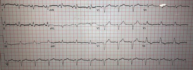
Here is his ED ECG:
 |
| What do you think? |
Both ECGs above show RBBB + LAFB, with massive concordant STE in leads V2-V6 as well as I and aVL. There is shark fin morphology (aka "Giant R wave", or "Lambda wave"), in which the wide complex QRS appears to fuse with the massive STE, causing confusion as to where the J point actually is, and where the QRS ends and the ST segment begins. This pattern is diagnostic of at least LAD occlusion (which of course supplies the anterolateral walls and the RBB and LAF), but given how severe the findings are even out into the high lateral wall, the occlusion may even be more proximal, such as the left main. This pattern is more commonly seen in LAD occlusion simply because persistent left main occlusion is rare, probably because it such patients rarely survive long enough to obtain an ECG and/or angiogram.
Below, I have placed red vertical lines at the J points.
I was sent the above ECG with no clinical information, and my response was: "Should be a huge LAD or theoretically even left main occlusion."
The cath lab was activated immediately. At this time the patient had stable ROSC, was intubated, and did not require pressors.
The LV EF was estimated at 20%. An Impella was placed for hemodynamic support.
Angiogram:
Acute thrombotic culprit lesion of the distal left main coronary artery and proximal LAD with 80% stenosis, also at the time of angiogram. There was also 80-90% stenosis of the proximal D1, and 90% at the ramus intermedius. It is not completely clear whether these were also thrombotic extensions of the distal left main lesion, but these are probably all part of the same acute culprit left main lesion, or downstream showering from it.
There was mild diffuse (non-quantified) CAD of the LCX, otherwise no evidence of CAD in any other part of the coronaries.
As predicted from the ability to obtain ROSC, there is not full occlusion at the time of cath. There is flow in each vessel distally.
For a patient who suffers cardiac arrest with recurrent or refractory VF due to left main occlusion, it is very unlikely to get stable ROSC unless the lesion opens up at least a little bit. And that is what we see here. The post cath ECG below confirms reperfusion including resolution of RBBB and LAFB:
In fact, now there is more of a LBBB pattern, but a bit odd because I and aVL are not all upright.Initial contemporary troponin T was 0.11 ng/dL (reference range less than 0.01 ng/dL). Troponin rose to 2.10 ng/dL but then no further measurements were recorded, so the peak troponin was not measured.
He had a long and rocky course in the ICU. Brain MRI showed multiple areas of restricted diffuse and acute cerebral infarct consistent with hypoxic encephalopathy. Despite ideal cardiac arrest conditions (immediate professional CPR, near the hospital, quick response time, seemingly zero no-flow time, and aggressive immediate cath lab activation), it seems that there will be at least some element of permanent neurologic injury, but the severity is yet to be determined.
Learning Points:
You must understand the shark fin morphology, and the fact that the LAD supplies the right bundle branch and the left anterior fascicle, if you want to be able to diagnose the most severe OMIs that exist like this one! STEMI criteria do not apply here, and many providers will not even know where to measure the ST segment at all.
In this case, we see lead aVR has ST and T wave depression in lead aVR, NOT ST elevation. This is because aVR simply shows the reciprocal findings of the many other leads which are oriented in the opposite direction. With widespread STE in many leftward leads, it is inevitable that lead aVR must show reciprocal STD. If there had been better coronary perfusion and more collateral circulation at the time of these ECGs, then instead of an anterolateral OMI pattern, we may have seen the pattern of diffuse subendocardial ischemia, with diffuse STD and reciprocal STE in aVR. The key is that aVR has no new or primary information. I teach that "aVR" stands for the "aVerage Reciprocal" lead, because it shows the reciprocal findings of the average of all the other leads on the 12-lead, which are largely oriented in the opposite general direction from lead aVR.
Sometimes even with the best possible conditions, the neurologic outcome of cardiac arrest can eclipse the cardiac salvageability.
Some of the most severe LAD or left main occlusions present with acute RBBB and LAFB, and these findings carry the highest risk for acute ventricular fibrillation, acute cardiogenic shock, and highest in-hospital mortality when studied by Widimsky et al. (in-hospital mortality was 18.8% for AMI with new RBBB alone). Additionally, the RBBB and LAFB make the recognition of the J-point and STE more difficult and more likely to be misinterpreted (when the QRS is wide, the J-point will hide!). Upon successful and timely reperfusion, the patient may regain function of the previously ischemic or stunned fascicles.
Widimsky PW, Rohác F, Stásek J, et al. Primary angioplasty in acute myocardial infarction with right bundle branch block: should new onset right bundle branch block be added to future guidelines as an indication for reperfusion therapy? Eur Heart J. 2012;33(1):86–95.
https://pubmed.ncbi.nlm.nih.gov/21890488/
See these other related posts:
A deadly alcohol binge: a man in his 30s with chest pain and initial high sensitivity troponin I within normal limits
See other "shark fin" morphology cases here:
Fascinating case of dynamic shark fin morphology - what is going on?
"Shark Fin": A Deadly ECG Sign that you Must Know!
Wide Complex Tachycardia; It's really sinus, RBBB + LAFB, and massive ST elevation
But don't be fooled by the other etiologies of shark fin morphology:


No comments:
Post a Comment
DEAR READER: I have loved receiving your comments, but I am no longer able to moderate them. Since the vast majority are SPAM, I need to moderate them all. Therefore, comments will rarely be published any more. So Sorry.