Written by Pendell Meyers with some edits by Steve Smith
A woman in her 60s on chemotherapy presented to the Emergency Department for a syncopal episode just prior to arrival. She was walking to the bathroom when she suddenly felt nauseous and passed out. EMS was called by the patient's daughter, and en route to the ED she vomited twice. On arrival to the ED, she adamantly denies chest pain but says she's "just still not feeling well." She had no prior known cardiac disease.
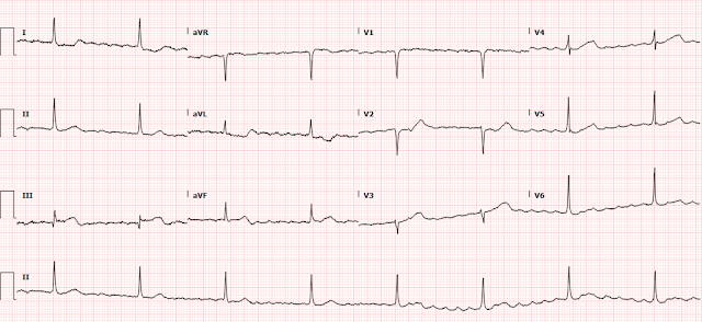
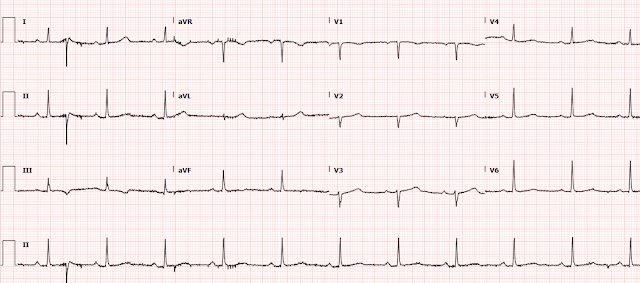
Triage at 0755:
The rhythm is most either atrial fibrillation with complete heart block and resulting junctional escape, or atrial flutter with very high degree (but constant) block. Remember, atrial fibrillation cannot result in regular ventricular rhythm if it is conducted through the AV node.
The QRS has narrow normal morphology. There is almost 1 mm STE in III, but no STE at all in II or aVF. The T wave in III is hyperacute, and is biphasic up-down. Leads aVL and I show reciprocal findings (STD and TWI biphasic down-up). Through the baseline wander, you can see the impression of downsloping STD in V2 indicating posterior extension. Interestingly, V3 then appears to have a hyperacute biphasic down-up T wave, with hyperacute Ts in V3 and V4, but then V5 and V6 do not. All the biphasic T-waves suggest that there is some early spontaneous reperfusion happening.
Overall, it is diagnostic of inferoposterior OMI, likely RCA occlusion, likely also explaining the acute bradydysrhythmia.
The rhythm was noticed as pathologic, but none of the ischemic findings discussed above were initially noticed. It seems as though the providers felt that the problem was a primary bradydysrhythmia.
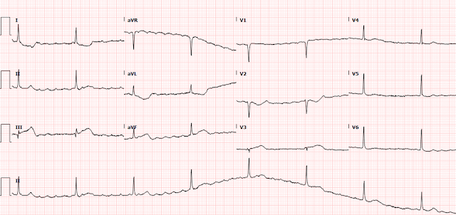
Initial troponin returned at 520 ng/L, and another ECG is recorded at that time (0900):
Findings are very similar to the first ECG. There is ongoing active transmural (full thickness) infarction. Still no STEMI criteria.
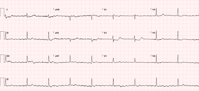
Anther ECG was done at 1000 (reason unknown):
This ECG shows some interval reperfusion compared to the prior ones, including slight deflation of the hyperacute T waves and terminal T wave inversion in III, and the opposite in V2.
At this point the patient was persistently hypotensive and bradycardic requiring epinephrine drip to maintain heart rate above 50 bpm. She was described as uncomfortable and short of breath. She continued to deny chest pain.
The ED team consulted cardiology for symptomatic bradycardia, asking whether the patient should go for an emergent pacemaker. The cardiology team evaluated the patient, reviewed all the ECGs above, and apparently found no concern for ischemia and agreed that the primary problem was just the bradydysrhythmia.
They were in the process of consenting the patient for a pacemaker when I contacted the ED provider in real time (I happened to be actively reading ECGs for the department from home at that time). I explained my concern for RCA occlusion as the cause of her OMI findings on ECG, and as the cause for her bradydysrhythmia (because the RCA almost always supplies the AV node).
The ED team performed a bedside echo which showed an inferior wall motion abnormality.
The repeat troponin I was 595 ng/L.
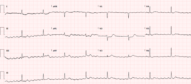
Another ECG was performed at 1055:
The end of the T wave in III is difficult to see through the atrial waves. There are some features of reperfusion, but also some leads that seem to show ongoing ischemia.
I was told the patient was still persistently symptomatic despite some features of reperfusion on ECG. She still required epinephrine and norepinephrine drip and was short of breath.
The ED provider was able to convince the skeptical cardiologist to perform an angiogram before placing a pacemaker.
The angiogram showed an acute thrombotic proximal RCA occlusion (TIMI 0 flow). After PCI, there was 0% residual stenosis with TIMI 3 flow.
Comment: TIMI-0 flow does not rule out some degree of microvascular reperfusion; collateral flow may account for the minimal ECG features of reperfusion. In fact, in Wellens' studies which established Wellens' syndrome, in all cases there was perfusion, but 20% of these cases were in spite of an occluded artery, but through collateral circulation.) This is precisely the reason why we conceptually define OMI as: "Acute coronary occlusion or near-occlusion with insufficient collateral circulation, resulting in imminent full-thickness myocardial infarction."
The first ECG after cath was performed hours later, in the cardiac ICU:
Sinus rhythm with PACs. There is resolution of the STE and hyperacute T waves, as well as terminal T wave inversion in III. This confirms reperfusion. Not to mention the dramatic improvement in AV node function.
The ICU staff noted that, after PCI, her heart rate rapidly improved and she never required even a temporary pacemaker. Her epinephrine and norepinephrine requirement resolved within hours of PCI.
In fact, within 24 hours the patient required metoprolol and amiodarone for rate control of intermittent atrial fibrillation with rapid ventricular response:
Later she was back in sinus rhythm:
She had several minor complications unrelated to ACS during her stay, but was ultimately discharged on day 5.
Learning Points:
The most important, deadly, reversible causes of bradycardia include acute RCA occlusion (OMI), hyperkalemia, and toxicity from beta blockers, calcium channel blockers, or digoxin. We in the ED must be the experts at recognizing these conditions on the ECG.
If this patient had instead received a pacemaker rather than RCA reperfusion, her ECG would have shown a ventricular paced rhythm. Providers who cannot recognize OMI in normal QRS conduction may be even less likely to recognize it in ventricular paced rhythm, although we have shown that it can be reliably done with proper training:
Until we get AI that can learn ECG patterns, we might need human ECG experts to act like radiologists, so that EM physicians and cardiologists can have access to expert interpretation.
You must learn to recognize hyperacute T waves.
This patient is STEMI(-) OMI that clearly benefits from emergent reperfusion.



No comments:
Post a Comment
DEAR READER: I have loved receiving your comments, but I am no longer able to moderate them. Since the vast majority are SPAM, I need to moderate them all. Therefore, comments will rarely be published any more. So Sorry.