Written by Pendell Meyers
A man in his 50s presented to the ED with chest pain described as pressure, without radiation, acute onset about three hours prior to arrival. He had had stuttering less severe versions of this pain all week that usually went away after a few minutes. He also had diaphoresis and dyspnea. He had extensive family history of CAD with CABG's in the mid 50s for multiple relatives, but he had no personal known history of CAD.
It is unclear whether he had pain at the time of triage, but notes describe that his pain had subsided by the time of EM physician evaluation:
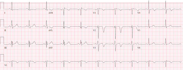
Triage at approximately 2130:
 |
| What do you think? Decide before viewing the baseline ECG available below, then decide again in the context of the baseline ECG. |
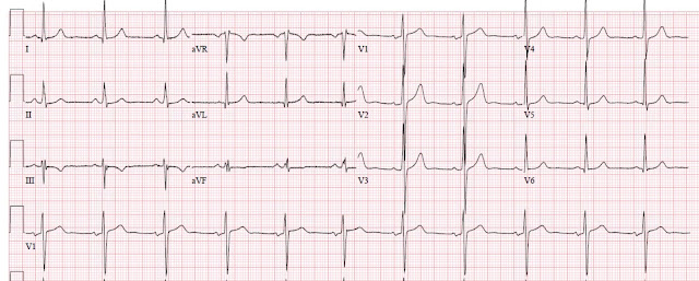
Baseline ECG on file from 5 years ago:
The baseline ECG is basically normal, with some high voltage possibly indicative of possible LVH (but no obvious LVH repol abnormalities), and borderline left axis deviation (also possibly due to LVH). These T waves and ST segments are normal for this QRS complex.
The triage ECG above shows reperfusion of OMI in the midanterolateral pattern which is now called "South African flag sign" (V2-3 areas, then "skips" the lateral precordial leads and reappears in I and aVL)
-- T-wave inversion in aVL, with flattening in lead I
--Reciprocally upright and hyperacute inferior T-waves (These appear to be inferior hyperacute T-waves, but are actually reciprocal to the inverted T-wave in aVL!!)
--Residual upsloping STE in V2 and V3, with terminal T-wave inversion. Important: "Wellens" pattern means reperfusion from OMI!
--There is also a new Q wave in V2, which must be considered pathologic until proven otherwise.
The pain was apparently resolved at the time of the first ECG.
The first troponin (contemporary -- NOT high sensitivity -- Troponin T, normal less than 0.01 ng/mL) was undetectable, reported at less than 0.01 ng/mL. In my 4-year experience with this particular assay, it does not even turn positive (detectable, 0.01 ng/mL or higher, which is also the 99% upper reference limit) until 4-6 hours after the event has started.
Another ECG was done around 2310, still without symptoms:
 |
| Not much change. Still appears reperfused. |
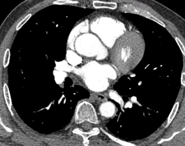
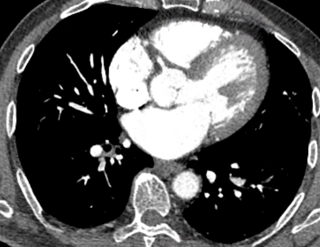
The providers ordered an emergent CT angiogram looking at both the aorta (for dissection) and a CT coronary angiogram (to evaluate for coronary stenoses or occlusion):
(Disclaimer: I have no training or expertise in interpreting CTCA, but I will give my best guess as to the arteries seen in the images below)
 |
| The left main bifurcation can be seen in the center of the heart in this image. |
 |
| In this image a small section of the LCX artery can be seen between the LV and the LA. |
 |
| Here we can see a section of the proximal and mid LAD, starting in the center of the heart and coursing to the upper right portion of the heart. We cannot fully evaluate the stenosis in this two-dimensional image, of course. |
 |
| In this image we can see the proximal RCA originating from the proximal aorta and coursing between the RV and RA. |
The emergent CT angiogram was read as:
No evidence of aortic dissection or pulmonary arterial thromboembolism.
Fair to poor quality due to significant calcifications.
Moderate stenosis in the mid LAD 50-69% associated with severe partially calcified plaques throughout the LAD. Please note that the degree of stenosis can be under or overestimated due to blooming and beam hardening artifacts which limits visualization of parts of the LAD in this case.
Minimal to mild multifocal stenosis due to partially calcified and noncalcified plaques in the LCX, left main, and RCA.
The second troponin was also undetectable, less than 0.01 ng/mL.
Another ECG was done around 0130:
 |
| Not much change. Still reperfused. Inferior T-waves are no longer reciprocally hyperacute |
The patient did well. No more troponins were measured. He was discharged the next day.
Learning Points:
Expert ECG interpretation often allows understanding of the exact state of the coronary lesion on arrival. It was subsequently confirmed in this case by both CT coronary angiogram and angiography.
Acute MI is not ruled out by contemporary troponins until AT LEAST 6 hours. (With high sensitivity troponin, a very small 1-, 2-, or 3- hour delta in the normal range can rule out MI with high negative predictive value. Contemporary troponins, especially Trop T, are not precise enough to use a delta below the 99th percentile)
In this particular case, I do not think coronary CT angiogram added much to the patient's management, as he was pain free and the ECG showed reperfusion. Perhaps, after two negative troponins, the team was starting to doubt ACS at all - perhaps this CT angiogram helped them stay focused on the correct diagnosis of ACS.
However, we believe there are several scenarios in which emergent coronary CT angio could be extremely helpful, such as:
A patient with suspected ACS and ongoing symptoms or ECG ischemia without STEMI criteria, who for whatever reason is not being taken for emergent angiogram due to lack of belief or understanding that the patient has OMI. In this case, if a CTCA were to show occlusion or near occlusion of a coronary vessel, in the context of acute symptoms, this may help the cardiologist understand the benefit to emergent management vs. delayed.
See this case where exactly that happened:
This patient with "NSTEMI" was not allowed to go to the cath lab. Then the ED provider obtained an emergent coronary CT angio. What do you think it showed?
A patient who has possible ACS symptoms and ECG abnormalities that are concerning but suspected to be due to a non-ACS cause, such as takotsubo cardiomyopathy, brugada pattern, etc. In such a case, emergent CTCA showing perfectly patent coronaries may help to avoid false positive cath lab activation. We don't currently have a recorded case demonstrating this use.
If you have used emergent CTCA to help diagnose or rule out OMI, thereby informing emergent management of possible ACS, please send us your cases!
See this study in which CTCA was fairly accurate for predicting angiogram findings in a population of NSTEMI patients (sadly they have no presented data on OMI, or acute lesions that would benefit from emergent rather than delayed NSTEMI treatment):
Management of MI can be similar to stroke: Use CT angiogram. Don't depend only on STE on ECG for reperfusion?
Here are cases where CT scan of the myocardium, seen incidentally on Chest/Abd/Pelvis CT, diagnosed OMI or NOMI:


No comments:
Post a Comment
DEAR READER: I have loved receiving your comments, but I am no longer able to moderate them. Since the vast majority are SPAM, I need to moderate them all. Therefore, comments will rarely be published any more. So Sorry.