Tuesday, February 4, 2025

Syncope and a short pause on event monitor

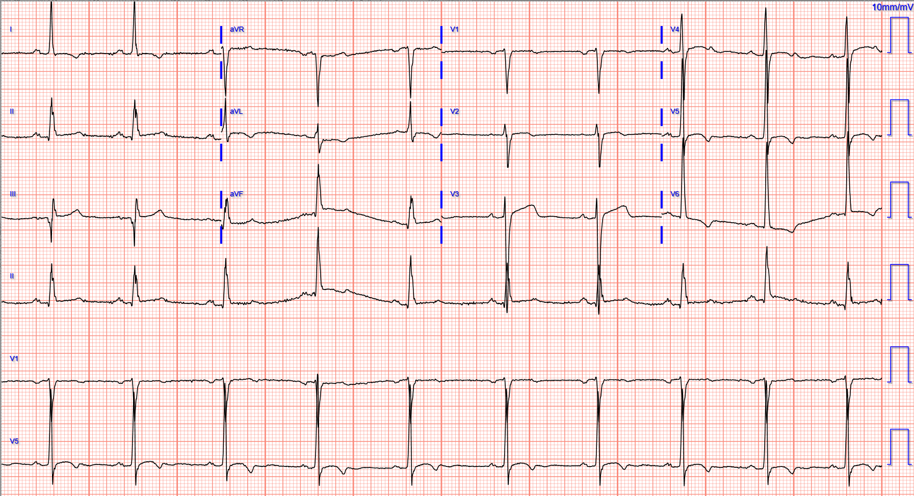
Written by Willy Frick

A middle aged man was inspecting the food in his (closed) oven when he felt a few moments of light-headedness. He subsequently woke up on the ground, unsure how long he had been unconscious. He presented for evaluation where inpatient workup including monitoring on telemetry was unremarkable. He was discharged with an event monitor and electrophysiology follow up.

The event monitor lasted for four weeks and showed almost no abnormalities, but the following strip was recorded.


What do you think?


Is the finding of just 3 blocked P waves on a 4 week event monitor enough to warrant permanent pacing?


Answer: YES. Remember that even a single episode of cardiac syncope at the wrong time (e.g. while driving) is enough to kill.

If the block is not vagal in nature, the patient should receive a pacemaker. Why do I make this distinction?

There was a randomized trial to determine whether pacemakers could benefit patients with vasovagal syncope, aptly named The North American Vasovagal Pacemaker Study (VPS). Patients with 6+ episodes of syncope and positive tilt table test were randomized 1:1 to pacemaker versus no pacemaker. What did they find?

(From VPS — Connolly et al  JACC, 1999 )

Major benefit to pacing! The relative risk of syncope was reduced by over 85%! The number needed to treat was 2. We almost never see benefit this profound! So...Why don't we pace patients with vasovagal syncope?

Answer: There was no sham control. Half the patients in VPS knew they had a fancy implanted computer to help the heart beat and prevent passing out. And half the patients knew they did NOT get such a device. Fortunately, this study was repeated with sham control. In the repeat study (VPS II), EVERY patient had an implanted pacemaker. Half of the pacemakers had pacing enabled, and half the pacemakers were set to sensing only without pacing. Most importantly, neither the patients nor the doctors knew which patients got which treatment, maintaining appropriate blinding. What did the authors find in VPS II?

(From VPS II — Connolly et al  JAMA, 2003 )

Answer: No significant reduction in syncope!! A stunning result. One of many examples in medical history that remind us of the importance of blinding in clinical trials. Why was there no benefit? Perhaps because the bradycardia in vasovagal syncope is only one part of the autonomic response. There is also systemic vasodilation resulting in transient hypotension and cerebral hypoperfusion. This is why we do not routinely pace patients with vasovagal syncope.

Back to the case:

Can we say any more about the cause of block here? Could it be vagal?


This is known as "paroxysmal atrioventricular block" due to a phenomenon called "phase 4 aberrancy." The short explanation is that a premature beat resets the His. The ensuing (very slight) prolongation in cycle length prolongs the duration of spontaneous diastolic depolarization in phase 4 of the action potential, rendering it refractory. Phase 4 block is also referred to as "bradycardia dependent block." It remains refractory until another extrasystole (escape beat, PVC, PAC, etc.) resets the His, allowing it to resume conduction. If you wish to read more about this, check out the references section at the bottom, especially the articles by Lee and Pillai. 

In the example above, the first PVC (boxed in red below) depolarizes the His at an unfavorable time rendering it refractory. Then three sinus P waves (black arrows) are blocked. Finally, a second ventricular extrasystole (boxed in blue) resets the His, and the next sinus P wave (orange arrow) marks the resumption of 1:1 AV conduction.


The way to recognize this type of block is that it both initiates and terminates with an extrasystole, and there is typically very little change in the underlying sinus rate. By contrast, vagal blocks are accompanied by slowing of the sinus rate.

The patient was given strict guidance not to drive, operate machinery, etc. and was scheduled for pacemaker implantation.

Learning points:
  • Diagnose phase 4 block, an indication for permanent pacing
  • Explain why we do not routinely pace patients with vasovagal syncope
  • Review VPS and VPS II, a historical lesson in the importance of sham control

References:

Connolly, S. J., Sheldon, R., Thorpe, K. E., Roberts, R. S., Ellenbogen, K. A., Wilkoff, B. L., Morillo, C., & Gent, M. (2003). Pacemaker therapy for prevention of syncope in patients with recurrent severe vasovagal syncope: Second Vasovagal Pacemaker Study (VPS II): a randomized trial. JAMA289(17), 2224. https://doi.org/10.1001/jama.289.17.2224 

Connolly, S., Sheldon, R., Roberts, R., & Gent, M. (1999). The North American Vasovagal Pacemaker Study (VPS). Journal of the American College of Cardiology33(1), 16–20. https://doi.org/10.1016/s0735-1097(98)00549-x 

Lee, S., Wellens, H. J. J., & Josephson, M. E. (2009). Paroxysmal atrioventricular block. Heart Rhythm6(8), 1229–1234. https://doi.org/10.1016/j.hrthm.2009.04.001 

Pillai, A., Ellenbogen, K. A., & Padala, S. K. (2021). A tale of 2 blocks. Circulation143(10), 1062–1065. https://doi.org/10.1161/circulationaha.120.052981 


===================================

MY Comment, by KEN GRAUER, MD (2/4/2025): 

===================================
Enlightening case by Dr. Frick — with lifesaving chance occurrence of a single short pause over a 4-week period of monitoring, without which the probable result would have been a preventable sudden (and unexplained) death of the patient.

Looking Closer:  The Single Short Pause ...
It is indeed uncanny that an otherwise negative 4-week period of monitoring captured such a "textbook picture" of PAVB (Paroxysmal AtrioVentricular Block) — in which we see both the onset of the several second ventricular pause precipitated by a slightly early ventricular beat (labeled "X" in my Figure-1) — as well as the offset of the pause prompted by a 2nd earlier-than-expected ventricular beat (labeled "Y" in my Figure-1).
  • As per Dr. Frick — the primary cause of one or more abrupt pauses with PAVB is a result of a diseased His-Purkinje system. That today's patient may be predisposed to this disorder is suggested in Figure-1 by: i) Obvious QRS widening on this single-lead ECG monitoring strip (suggesting some form of bundle branch block); — andii) 1st-Degree AV Block of conducted beats (PR interval ~0.26 second).
  • Although the continuous rhythm strip in Figure-1 does not manifest slowing of the atrial rate prior to onset of the ventricular pause (ie, the atrial rate is fairly constant at ~66/minute in the first 2 lines of the rhythm strip) — there is a change in the atrial rate after the return of 1:1 AV conduction (in the form of slight acceleration of the atrial rate to ~81/minute for 6 beats — until resumption of the slower atrial rate at the end of the 3rd line). So, perhaps a component of vagal influence may be operative after all? (ie, 1st-degree AV block — and some rate variability).
  • "Devil's Advocate" — Beat "Y" actually looks like a fusion beat (intermediate in morphology between sinus-conducted beats and early ventricular beat "X" ) — which if true, would make for an immediate "reset" of His-Purkinje conduction.

Figure-1: I've labeled today's continuous rhythm strip.


Paroxysmal AtrioVentricular Block (PAVB):
As suggested by Dr. Frick's discussion — and described by Bansal et al (J Arrhythmia 35:870-872, 2019) and Bosah et al (Cureus 14[7]: e27092, 2022) — the entity known as PAVB is a potential cause of syncope that is easily overlooked, and which is potentially lethal (probably more often than is commonly realized).
  • PAVB is characterized by the sudden, unexpected onset of complete AV block with delayed ventricular escape — therefore resulting in a potentially prolonged period without any QRS on ECG. Prior to the prolonged pause — the patient manifests 1:1 AV conduction without other evidence of AV block (which is why onset of PAVB is typically so unsuspected! ).
  • Because of its totally unexpected onset and propensity to result in sudden death — PAVB is difficult to document and significantly underdiagnosed.
  • Three mechanisms for producing PAVB have been described: i) Vagally mediated (ie, Vagotonic Block — as described in the January 31, 2019 post, — and in more detail HERE — with the references listed at the end this Blog post citing instances of transient asystole from excessive vagal tone!)ii) Intrinsic (Phase 4 = pause- or bradycardic-dependent) PAVB; — andiii) Idiopathic.

i) Vagotonic AV Block:
This form of PAVB is potentially benign when it results from a transient profound surge of parasympathetic tone in an otherwise healthy individual (as might occur with an episode of severe vomiting; a fit of severe coughing; vasovagal reaction from a blood draw).
  • The problem with vagotonic PAVB is localized to within the AV Node.
  • There will often be a "prodome" of diaphoresis, nausea, dizziness — with the patient aware of imminent fainting.
  • Characteristic ECG findings of vagotonic PAVB include progressive sinus rate slowing — often associated with an increasing PR interval and a narrow-QRS escape focus — followed by recovery with progressive return to a normal sinus rate and normal PR interval.

ii) Intrinsic PAVB:
Several names have been attached to this mechanism of PAVB — including most commonly "Phase 4 AV block" and/or PD-PAVB (Pause-Dependent PAVB).
  • PD-PAVB appears to be the primary mechanism for the cardiac rhythm in today's case (albeit potential for some vagal contribution given variability in the atrial rate in Figure-1). As per Dr. Frick — the underlying pathology of PD-PAVB is severe His-Purkinje System disease (supported by the finding of QRS widening with conducted beats in today's rhythm strip). This form of PD-PAVB is likely to be fatal unless the patient receives a permanent pacemaker.
  • As is superbly shown by Dr. Frick's case — the interesting pathophysiology of PD-PAVB results from chance occurrence of an "appropriately-timed" PAC or PVC that partially depolarizes the diseased HPS (His-Purkinje System) at a specific point in the cycle that renders the poorly-functioning HPS unable to complete depolarization. The resultant prolonged pause in ventricular depolarization may only resolve if another "appropriately-timed" PAC or PVC occurs at the precise point needed to "reset" the HPS depolarization cycle (which presumably explains why the patient in today's case spontaneously recovered).
  • Of note — although QRS widening and 1st-degree AV block is seen for conducted beats in today's case — up to 1/3 of patients with PD-PAVB do not show evidence of conduction defects on ECG. This complicates documentation and explains why it is so easy to miss the diagnosis.

iii) Idiopathic PAVB: 
This is the most recently described form of PAVB — in which findings are not fully consistent with either of the other 2 forms.
  • The baseline ECG before idiopathic PAVB tends to be normal.
  • No "trigger" for PAVB is evident (ie, no source of excessive vagal tone — and no precipitating PACs/PVCs are seen).

============================
BOTTOM Line: It's important to be aware of the various forms of PAVB — that up until recently were all-too-often undetected, especially in patients without evidence of conduction defect on their baseline ECG.
  • It is fortunate that this patient was monitored over an extended period of time ( = 4 weeks). It is humbling for me to see how easily the diagnosis could have been missed in today's patient.


Sunday, February 2, 2025

Sometimes a patient is fortunate to have a cardiac arrest

Written by Pendell Meyers 


A man in his 60s presented with acute chest pain.

Here is his triage ECG:

What do you think?


There is sinus rhythm with clear LVH. Leads V5-6 are suspicious for upright, enlarged T waves that are possibly inappropriate for the QRS complex, especially V6. But without a baseline for comparison, it would be difficult for me to say that it is specific and diagnostic for OMI. The Queen of Hearts agrees, calling the ECG not specific for OMI.

Smith comment: V6 is very atypical for LVH and very suspicious for OMI.  There is a very high voltage R-wave without any S-wave (terminal QRS distortion) and with a concordant ST segment and T-wave, with convex ST segment.  It is essential to compare with old ECGs.  In the absence of old ECGs, one must record serial ECGs every 15 minutes.

He had several older ECGs on file, here are two examples:

6 days prior:


2 months prior:


In the context of ACS symptoms, and when able to compare the new vs. old ECG, the top ECG is DIAGNOSTIC of OMI until proven otherwise. 

Unfortunately, the ECG was interpreted as no significant change from prior, "no STEMI"!!

He was sent back to the waiting room, where he suffered a VF arrest. Defibrillation was performed, and ROSC was achieved. 

Approximately 5 minutes after ROSC, this ECG was obtained (about 45 minutes after arrival):

Obvious anterolateral OMI, and STEMI criteria positive for those who care or need it.

Smith comment: The patient was lucky to have a cardiac arrest.  Had he not had one, he would have sat in the waiting room until his entire myocardium at risk infarcted.  By undergoing an arrest, providers became aware of his OMI which had not been noticed on his diagnostic ECG, and he thus has a chance at some myocardial salvage.

Also: notice how the LAD Occlusion results in DECREASED S-wave amplitude, compared to the previous ECGs, in V2 and V3.  I have noticed that this nearly always happens, and is the reason that you rarely find an ECG that shows BOTH LVH with high right precordial amplitudes AND acute STEMI/OMI.


Total proximal LAD occlusion was found and stented at angiography soon after the ECG above. Here are some images:


Next morning ECG:
Reperfusion findings are clear..
There is some R-wave preservation, a good sign that some myocardium remains viable


Echocardiogram:
EF 15%
Severe global hypokinesis
Akinesis of basal-mid inferior and inferoseptal myocardium
Severe hypokinesis to akinesis of apical anteroseptal, anterior, and inferoseptal myocardium

An echo from 3 months ago showed an EF of 40% with global mild hypokinesis, without focal wall motion abnormalities.

Smith comment: the initial echo was so bad because even myocardium that was salvaged was "stunned" and it takes time to recover function.  Weeks later, the EF improved from 15% to 40%.  If this patient had not had ventricular fibrillation and thus not had a reasonably short door to balloon time, all of that stunned myocardium would instead be permanently infarcted, with permanent 15% EF.

Troponin I quickly exceeded the laboratory limit of reporting at 25,000 ng/L.


4 days later:

Continued reperfusion.


He was discharged home, but quickly bounced back 4 times within the next few months for worsening heart failure. Long term outcome is not available.


===================================
MY Comment, by KEN GRAUER, MD (2/2/2025):
===================================
We need to learn from cases like today's. Today's initial ECG is not an easy tracing to interpret. This is because (as per Dr. Meyers) — there is obvious LVH that complicates assessment for potential acute ST-T wave abnormalities.

  • To Emphasize: Once again — the history is KEY. This patient is a man in his 60s who presented to the ED with new CP. As a result — without yet looking at the initial ECG, our "mindset" has-to-be awareness that this history alone places this patient in a higher risk category for having an acute cardiac event. 
  • As a result — Our "mindset" has-to-be that we need to rule out an acute event, rather than the other way around. This means that if there is any uncertainty about the diagnosis (or about the initial ECG) — that we need to "dig further" until we arrive at more certainty in our diagnosis. Unfortunately, this was not done in today's case. I say this — because compared to an ECG done just 6 days earlier — there clearly has been significant ST-T wave change, that in this patient who presents to the ED with new CP — has-to-be recogized as an acute OMI until proven otherwise (See Figure-1 below — in which I place these first 2 tracings in today's case together).

What Are the Oversights?
In my experience — many (if not most) clinicians do not compare serial ECGs by placing both ECGs that they are looking at side-by-side.
  • I fully acknowledge that I miss important findings when I do not do this. I know this was not done in today's case — because if side-by-side comparison had been done — then the difference in ST-T wave appearance between leads V4,V5,V6 in ECG #1 and ECG #2 would not have gone unnoticed. 
  • We have often remarked on the challenge that LVH may pose for recognizing acute ischemic changes. This is because LV "strain" often manifests straightened and elevated ST segments in anterior leads with deep S waves from marked LVH. For example, as an isolated finding — I would not necessarily interpret the ST segment straightening and elevation that we see in leads V2,V3 of ECG #1 (outlined in RED and light BLUEas abnormal in a patient with marked LVH.
  • The other challenge posed by the ECG of a patient with marked LVH with "strain" — is distinguishing between the ST-T wave inversion in one or more lateral leads due solely to LVH — vs that due to acute ischemia or infarction. This distinction is further complicated because marked LVH may at times mask the ST-T wave changes of acute ischemia. That said — the asymmetric ST-T wave depression in lead V6 of ECG #2 is classic for the ECG appearance of LV "stain". But 6 days later (ie, when ECG #1 was recorded) — there is not even a hint of ST flattening or depression in leads V4,V5,V6. This means that the ST-T wave appearance in ECG #1 clearly represents an acute change (compared to ECG #2) that would not be missed if there had been lead-to-lead comparison between these 2 tracings.

Figure-1: Comparison between today's initial ECG — with a prior ECG done just 6 days earlier.


How to Remember ECG Findings of LVH and LV "Strain"?

There are over 50 criteria in the literature for the ECG diagnosis of LVH. And — the ST-T wave appearance of LV "Strain" and "Strain Equivalents" is subjective, and can be confusing.

  • To facilitate recall of these concepts — We've conveniently added a link in the menu at the top of every page in Dr. Smith's ECG Blog that takes you to My Comment in the June 20, 2020 post, in which I review a user-friendly approach to ECG recognition of LVH and "Strain" (See Figure-2).


Figure-2: Handy link to LVH Criteria (conveniently placed in the menu at the top of every page in Dr. Smith's ECG Blog).


WHY then is Today's Initial ECG So Difficult to Interpret?

The above said — I still found today's ECG incredibly difficult to interpret for the simple reason that there is marked overlap of QRS complexes in virtually all of the chest leads.

  • Confession: I spent over half an hour staring at the QRS complexes in each of the chest leads, trying to distinguish between the limit of R waves from overlapping S waves. I am still not certain that my color-coding in Figure-1 is completely correct.
  • KEY Point: Whereas the ST segment straightening and elevation that we see in leads V2 and V3 of ECG #1 does not necessarily look abnormal (because as noted earlier — LV "strain" from marked LVH may show this ST-T wave appearance in anterior leads) — the same is not true for leads V4,V5,V6.
  • Lead V4 in ECG #1 (outlined in dark BLUE) — manifests an S wave that is only 13 mm deep. Doesn't the coved and elevated ST segment in this lead look disproportionate and abnormal?
  • As noted earlier — LV "Strain" manifests as ST-T wave depression in lateral leads (or at least as ST-T wave flattening). LV "strain" does not manifest as ST segment coving and elevation, as we see in lead V5 and especially in lead V6 of ECG #1.


What Could Have Been Done to Facilitate Interpretation? 

  • Rather than normal standardization — I would have immediately repeated today's initial ECG at half-standard voltage. Doing so would have minimized overlap — and facilitated appreciation of disproportionality by clarifying relative size of QRS complexes and ST-T waves.


Recommended Resources