Wednesday, September 29, 2021

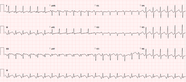
Massive ST Elevation in a 38 year old with Syncope

This ECG was texted by a former resident with the words "38 year old, syncope while urinating.  Negative troponin."

What do you think?


There is massive ST Elevation of 5 mm (at the J-point, relative to the PQ junction) in lead V2.  There is 3 mm in lead V1 and 2.5 in lead V3.  

But there is also 57 mm QRS in V2 and a 19 mm R-wave in V4.  The QT interval is not very long.

My response was this: "I have seen this pattern before and it is very unlikely to be OMI."  I did not know what "troponin negative" referred to [single troponin?  below the level of detection?  2 serially negative trops below the LoD?  Or below the 99th percentile.]  Nevertheless, I added: "And if you now have negative serial troponins, you have proven it."

Clearly the provider who sent this to me strongly suspected it was a false positive (as he/she did not act on it except to measure troponin), and I don't know if that is due to low pretest probability, patient appearance/exam, or knowledge of this PseudoSTEMI pattern.

Can we use the formula?

12 Example Cases of Use of 3- and 4-variable formulas, plus Simplified Formula, to differentiate normal STE from subtle LAD occlusion

First, we must answer all these questions in the negative:

1. Is there BBB?

2. Is there T-wave inversion?

3. Is the ST segment more than 5 mm in any lead (here it = 5 mm)

4. Is there terminal QRS distortion?

5. Do any of the leads with ST Elevation have a convex ST segment (here they are straight, which is also worrisome, but they are not convex)

6. Is there significant ST depression in inferior leads

7. Is there STD in V2-V6?

8. Are the pathologic Q-waves in any of V2-V4?

So, yes, we can use it.

If we use the 4-Variable formula, we get: corrected QT interval of 400 msec, QRS V2 of 57 mm, R wave in V4 of 18.5 mm, and ST Elevation at 60 ms after the J point in lead V3 = 4.5 mm.

This gives a very low value of 12.0.  

No result, no matter how low, absolutely rules out LAD occlusion, and if your suspicion is high, then go with your clinical impression.  The formula is mostly to be used when you DON'T think there is OMI.  It is not to dissuade you from OMI!!

But this is a case of syncope, not chest pain.  So the pretest probability is much lower.  And I'm telling you to memorize this morphology as one that is occasionally seen in non-ischemic ECGs.

In such a case, it is OK to delay just a few minutes to find an old ECG, or establish absence of wall motion abnormality.

It is also OK to have a false positive cath lab activation.

This patient would not stay for further workup, so whether there was structural abnormality or not is uncertain. But I can say that I have seen normal variants with this appearance.

Thursday, September 23, 2021

Cardiac Arrest at the airport, with an easy but important ECG for everyone to recognize

 Written by Pendell Meyers


Every once in a while we need to go back and cover some easy but important ECGs.

This will be far too easy for most readers of this blog, so please go find a learner and show them this case. Make sure they understand this case well, so that they will be able to learn from the harder versions of this case.


A middle aged female suffered sudden witnessed cardiac arrest at the airport, with quick bystander CPR.

EMS arrived and found her in VF. She was successfully defibrillated.


Her EMS ECG on the way to the ED was sent to us:


What do you think?


There is likely sinus tachycardia with a prolonged PR interval. Some learners might be worried about VT initially, if they do not recognize the QRS. Instead it is RBBB and LAFB. There is striking concordant STE in V2-V4 that is easy and unmistakable in this case, but often times is much harder to recognize when the J point is not quite as pronounced as it is in this case (see harder cases below).


This pattern is one of the highest risk OMI patterns recordable on an ECG: it is large proximal LAD occlusion until proven otherwise. The LAD supplies the anterior septum where the right bundle branch and left anterior fascicle are located. Acute proximal LAD occlusion can cause ischemia of these fascicles causing the combination of RBBB and LAFB. See below for review and references.


The cath lab was activated, and the patient fortunately suffered no further arrests. She was of course found to have an ostial LAD occlusion that was opened and stented.

The initial high sensitivity troponin I resulted elevated at 105 ng/L.

Here is her ECG the next morning:

The RBB and LAF have recovered. But there is significant loss of R waves anteriorly, with QS waves and some mild persistent STE. There has been a very large area of irreversible infarction.


The second troponin was around 16,000 ng/L, and no further troponins were ordered.

She survived the hospitalization.


Learning Points:

Like LBBB and many other abnormal QRS patterns, the formal "STEMI criteria" do not apply to RBBB with LAFB. There are no formal criteria for this situation, nor any "STEMI equivalent" criteria in the 4th universal diagnosis of MI. In standard 2021 practice, what generally happens is: if the STE is striking and clear like it is in this case, everyone understands it is "STEMI". If the STE is not as striking, or the J point is more obscured by the QRS pattern than in this case, it is often not recognized and missed. See the more difficult cases below.


Some of the most severe LAD or left main occlusions present with acute RBBB and LAFB, and these findings carry the highest risk for acute ventricular fibrillation, acute cardiogenic shock, and highest in-hospital mortality when studied by Widimsky et al. (in-hospital mortality was 18.8% for AMI with new RBBB alone). Additionally, the RBBB and LAFB make the recognition of the J-point and STE more difficult and more likely to be misinterpreted. Upon successful and timely reperfusion, the patient may regain function of the previously ischemic or stunned fascicles.


Widimsky PW, Rohác F, Stásek J, et al. Primary angioplasty in acute myocardial infarction with right bundle branch block: should new onset right bundle branch block be added to future guidelines as an indication for reperfusion therapy? Eur Heart J. 2012;33(1):86–95. 

https://pubmed.ncbi.nlm.nih.gov/21890488/

This pattern of LAD OMI with RBBB and LAFB is so high risk, I would estimate in my experience at least 20-50% of these patients suffer severe cardiogenic shock or arrest before emergent PCI can even be performed. 

See these examples below, almost all of which are more difficult than this case:

A woman in her 60s with 6 hours of chest pain, dyspnea, tachycardia, and hypoxemia


Tuesday, September 21, 2021

A woman in her 60s with palpitations, chest discomfort, and multiple misdiagnoses by both EM and Cardiology!!

 Written by Pendell Meyers


A woman in her 60s was shopping when she suddenly experienced palpitations and chest "discomfort." She denied outright chest pain or dyspnea. She walked across to the street to my Emergency Department. She had no known prior history of dysrhythmias or heart disease, but had known hypertension, breast cancer, diabetes, and obesity. She has had episodes of palpitations in the past, followed by holter monitor workups which did not reveal any cause of palpitations. However, her symptoms today feel worse than prior episodes, and she has never felt the "chest discomfort" with prior palpitations. Upon pointed questioning, she told the providers she has had several similar episodes over the past few weeks, but did not seek care during those episodes and they were shorter.


Here is her triage ECG during active persistent symptoms:

What do you think?


We see a regular, narrow, monomorphic tachycardia, for which the full differential would include sinus tachycardia, SVT (an umbrella term including many different rhythms), and atrial flutter. 

This ECG has a large negative atrial wave just before the QRS complexes in the inferior leads, with only one of these waves visible for each QRS complex. These waves are of course fully upright in V1. The differential includes a low ectopic atrial tachycardia near the AV node, or a relatively high AVNRT such that the circuit activates the atrial retrogradely before the circuit can activate the ventricles anterogradely during each lap of the circuit.

It is not atrial flutter, because with such a prominent atrial wave seen in the inferior leads we should be able to see a second set of these waves midway between the visible ones.

Whatever the atrial waves are, their QRS complexes in the inferior leads are followed by ST segments which seem to be slightly above the baseline in III and aVF. Because I have been fooled by this phenomenon before, and because it does not match OMI patterns I've seen before, I can see that this morphology is likely due to the upright atrial repolarization wave from the dysrhythmia. If there were any question whether it were due to OMI, I would first convert the patient out of this dysrhythmia then reevaluate the ECG for OMI.


The Emergency Medicine team perceived inferior STE in II, III, and aVF, with slight reciprocal STD in aVL, and decided to activate the cath lab for chest discomfort and possible STEMI. Cardiology came immediately and deactivated the cath lab after looking at this ECG and believing that it represented atrial flutter with 2:1 block (this is almost certainly not atrial flutter). 


Approximately 20 minutes later it spontaneously converted to sinus rhythm:


2 hours later it recurred:


She was given 10 mg of diltiazem IV and had conversion back to sinus soon afterward.


Prior ECG on file:


Two high sensitivity troponin I measurements were less than 6 ng/L.


The cardiology team incorrectly diagnosed this rhythm as atrial flutter with 2:1 block.

She was admitted for rate control and "new onset atrial flutter workup."

Echo was normal.

Unfortunately, in the mindset of atrial flutter, the patients CHADS2VASc score was calculated at 3, and she was deemed to require anticoagulation with eliquis (apixaban), which she has been on ever since (several months at this point). I am trying to contact her providers to discuss this.

She was also placed on metoprolol, presumably for rate control during her perceived atrial flutter episodes. In reality, it may be suppressing her SVT (whether it is ectopic atrial tachycardia or AVNRT).


Check out these posts showing atrial repolarization (Ta wave) and how it can cause inferior pseudoSTEMI/OMI patterns:

A man in his sixties with chest pain


Look at this ST Depression (It's not real)


Atrial Repolarization wave mimicking ST Elevation:

Sudden CP and SOB with Inferior ST Elevation and in STE in V1. Is it inferior and RV OMI?

A man in his sixties with chest pain

Learning Points: 

Atrial repolarization can be especially visible in ectopic atrial tachycardias (or any dysrhythmia with pronounced ectopic atrial waves), causing the appearance of inferior STE that has nothing to do with OMI.

Diltiazem should not generally cause conversion of atrial flutter to sinus rhythm. Diltiazem converting the rhythm would be more consistent with a reentrant dysrhythmia involving the AV node, such as AVNRT. There is always a chance that the conversion was actually just spontaneous and not associated with the diltiazem.

Misdiagnosis of atrial flutter can lead to significant long term patient harm, especially when treated with anticoagulation. Sometimes getting the rhythm correct is very important. 


Sunday, September 19, 2021

A man in his 50s with anterior ST elevation and a "tall T wave in V1"

Written by Pendell Meyers


Take a look at this ECG from a 57 yo M without any context first:


What do you think? Imagine he presented with chest pain.


There is normal sinus rhythm. QRS shows high voltage, likely representing LVH. There is STE in V1-V4 measuring up to 3-3.5 mm, and STD in V5-6. V2 has saddleback morphology, and V3 has a straight ST segment. V4 has slightly convex ST segment.

Findings that would potentially favor OMI: large absolute amount of STE, large proportion of STE to QRS in V1-2, STD in areas like V5-6 that would be considered reciprocal to V1-2 area, straight ST segment in V3 and slightly convex in V4.

Findings that would potentially not favor OMI: high voltage, LVH, saddleback morphology in V2, LVH with appropriate discordant ST/T segments explaining STE in right precordial leads and STD in left precordial leads. Saddleback morphology in V2, when in the same location as the STE that you are potentially worried about, is a quite strong feature suggesting a pseudoSTEMI pattern rather than true OMI.

In the proper clinical context, it would not be wrong to call this concerning for STEMI(+) OMI. But with more and more experience, some of these false positive features can be distinguished from true positives. Regardless, the first things to check would be the clinical context and any prior ECGs if available.


This ECG was sent to me without any context, and I responded, "I see why you are concerned, but I think this is likely a false positive from LVH. I would not activate the cath lab for this ECG at this point, but tell me more about the clinical picture and any prior ECG if available."

I sent it to Dr. Smith with no context, and he immediately responded: "very interesting pseudoSTEMI pattern"


In this case, the patient had only syncope which was explained by alcohol intoxication, poor PO intake otherwise, and standing in a line for a prolonged period of time. He did not have any active ACS symptoms. 

Additionally, he was fortunate enough to go to an ED where they had his prior ECGs on file: 


One of the clinicians who saw this patient told me they were worried because of "new tall T wave in V1." They stated that the prior T waves in V1 were flat or had terminal inversion. The present ECG above has a fully upright and larger T wave. That is true, but there is something about the morphology of it that does not match the many prior hyperacute T waves I have seen in lead V1. It comes with experience, and with seeing the other pseudoSTEMI features in this case.


His troponin was less than 6 ng/L, and other basic labs within normal limits.

He had a prior echo on file showing symmetric LVH which was attributed to longstanding hypertension. 

He had no family history of sudden death or obvious known HOCM, Brugada. 

He was observed for sobriety and no further episodes of syncope or cardiac dysrhythmias were noted.

He was discharged.


See these examples of saddleback morphology in V2:

Saddleback ST Elevation. Is it STEMI? Is it type II Brugada?


See these other great and related posts about LVH:

Syncope, History of Coronary Disease, and ST Elevation: Should Medics Activate the Cath Lab?


Profound ST Elevation in V1-V3. What do you think?


A 31 year old with Diabetes and HTN complains of bilateral arm tingling and headache


Huge Anterior Voltage, with ST Elevation


Hyperacute T-waves? Anterior STEMI? No, LVH with PseudoSTEMI pattern!


Learning Points:

LVH causes some of the most common and most difficult ECG patterns to differentiate from STEMI and OMI.

When LVH causes pseudostemi patterns, the STE that catches everyones eyes is usually in the right precordial leads V1-V3, where it is discordant to the QRS of LVH.

Saddleback morphology, often in or around lead V2, is very uncommon in OMI of that same location, to the point that we have only been able to report 2 such cases (see links above), despite 1,326 (!) published as of today.

ST segment convexity and straightening are features that somewhat favor OMI, but this is most applicable in a normal QRS complex context.

Comparison to prior ECGs and the clinical context are obviously and always paramount, and can frequently help you out of false positive STEMI criteria scenarios like this.


Friday, September 17, 2021

Narrow Complex Tachycardia at a Rate of 220

40-something yo who is on flecainide and diltiazem had sudden onset chest pain, palpitations, shortness of breath and diaphoresis:

Rate is 220.  What do you think?


It is fast, narrow, and regular, without P-waves.  So it is not atrial fib and not VT.   It is a regular narrow complex tachycardia.   There is a lot of ST depression -- this is ischemia caused by the very fast rate and is an indication for emergent electrical cardioversion.

What is the DDx?  

----PSVT (which includes AVNRT and orthodromic AV reciprocating tachycardia) [AVRT uses an accessory pathway, a "bypass tract" and thus is seen in WPW; about 30% of PSVT is orthodromic AVRT] 

----Atrial Flutter with 1:1 conduction.

Which is it?

The fact that the patient is on Flecainide and Diltiazem is good evidence that this is atrial flutter with 1:1 conduction.  These medications are primarily given to patients with atrial fib or flutter (the flecainide keeps the patient in sinus rhythm, and the diltiazem prevents 1:1 AV conduction when the patient is in flutter).

If you look closely at lead II across the bottom, it appears there are flutter waves.

This is atrial flutter with 1:1 conduction.  The patient is not on anticoagulants, but the very fact that he is on flecainide is evidence that he probably spends most time in sinus rhythm.

The providers thought this was AVNRT.

He was given adenosine 6mg, then 12mg, with no change.  If AVNRT, then one would expect conversion.  If flutter, one would expect AV blockade and uncovering of obvious flutter waves.  But the dose of adenosine clearly was not high enough to sufficiently block the AV node.  One could escalate to 18 mg.  This was not done.

Instead, they appropriately decided on electrical cardioversion.  The decided to sedate with etomidate. After giving the etomidate, but before cardioversion, they noticed that the patient had "converted":

Here is the 12-lead:

What happened?  Rate is now 120.
Ischemia is gone.

The providers thought that the patient had spontaneously converted.  

However, look closely (this explanation is fascinating -- pay attention!!): there are still flutter waves.  The flutter rate is 240 and there is 2:1 conduction with a ventricular rate of 120.  Go back to the first ECG, and the ventricular rate was 220, which means that the atrial flutter rate has INCREASED from 220 to 240!!

The reason that the ventricular rate has slowed is that the flutter rate has increased!!

The AV node cannot conduct as a rate of 240, but it can conduct at a rate of 220.  When the atrial flutter rate increased, the AV node could not keep up and the patient got much better because of a decreased ventricular rate.

Why did the flutter rate increase?  Not certain, but just a change in sympathetic tone would do it.

A short time Later the patient actually converted to sinus:


What would have a happened with a higher dose of adenosine?  The AV node would have blocked the conduction for a brief time and the flutter waves would have become obvious. Then the adenosine would be quickly metabolized, and the AV node would resume 1:1 conduction.  So adenosine does not treat this, but sometimes makes the diagnosis obvious.


Learning Points:

1. Patients who get atrial flutter and are kept in sinus rhythm with a type I antidysrhythmic, usually flecainide, must be on an AV nodal blocker to prevent 1:1 conduction.  In this case, it is uncertain if he had not taken his diltiazem, or if he needed a higher dose.

2. Regular Narrow complex tachycardia, if not sinus tach, is AVNRT, AVRT, or atrial flutter with 1:1 or 2:1 conduction.

3.  Never try to convert atrial flutter with a drug such as procainamide without first giving an AV nodal blocker.  You will cause a slowing of the atrial flutter rate with potential to INCREASE the ventricular rate.


Thursday, September 16, 2021

Syncope in a young man

 Written by Pendell Meyers


Let's say a young person presented with exertional syncope. They are now at baseline, asymptomatic, normal vital signs, and they have this ECG at triage:


What do you think?


Below are two other variations of this patient's resting ECG from different time periods:


Answer: Arrhythmogenic Right Ventricular Cardiomyopathy

See the end of the post for review and details on ARVC and it's ECG findings, but suffice to say that this patient has sinus rhythm, small epsilon wave in some of his ECGs, and R waves in V1-V3 with TWI.


Here is a close up of V1-V3:

Notice the very subtle micro-voltages at the J point.  These are epsilon waves.


As a teenager, this patient was playing basketball when he suddenly collapsed and became unresponsive. EMS found him in ventricular fibrillation. He returned neurologically intact. During his hospitalization he received an ICD. Genetic testing confirmed arrhythmogenic right ventricular cardiomyopathy/dysplasia with a mutation in his desmoglein-2 gene. During hospitalization he was started in mexiletine for frequent uniform PVCs which seemed to significantly decrease ectopy burden. He was restricted from activity and later also metoprolol was added.

Over the years he has had several ICD shocks, several medication adjustments, but is otherwise doing well. 


ARVD Review below, reproduced from this post: 

Young man with syncope while riding a bike [Arrhythmogenic Right Ventricular Dysplasia (ARVD)]


ARVD, also known as arrhythmogenic RV cardiomyopathy, is estimated to have a prevalence of 1 in 5000 adults and is responsible for approximately 11% of sudden death in young adults and 22% in a study of athletes in northern Italy.  The diagnosis is not easy (see below).

There is a 2010 publication by the Task Force in Diagnosis of ARVD: Diagnosis of arrhythmogenic right ventricular cardiomyopathy/dysplasia: proposed modification of the task force criteria. 

There are 6 categories of criteria
1) Imaging
2) Pathologic
3) ECG Repolarization
4) ECG Depolarization
5) Arrhythmias
6) Family History.  

ECG and historical Highlights of this publication are (Suspect ARVD with):
1) High risk syncope with no other etiology; Family History
2) Depolarization abnormalities (Major criteria):
        a) Epsilon Waves
        b) Localized prolongation (greater than 110 ms) of the QRS complex in right precordial leads (V1-V3)
3) Repolarization abnormalities in patients of age at least 14 years (because younger patients often have juvenile T-waves)
        a) Minor: Inverted T-waves in right precordial leads V1-V2 
        b) Major: Inverted T-waves in right precordial leads V1-V3 or beyond (major criteria) 
4) Arrhythmias
        a) Major criterion:
                i) VT of LBBB morphology with superior axis (negative or indeterminate QRS in leads II, III, aVF and positive in lead aVL) (major criteria)
        b) Minor criteria:
                i) VT of LBBB morphology with inferior axis (positive QRS in leads II, III, aVF and negative in lead aVL) (minor criteria)
                ii) More than 500 PVCs per hour
5) Finally, it is a progressive disease and patients without ECG abnormalities may develop them over time.


Here is an example of an epsilon wave (image C).  And another example.  Here are some great examples from the post on RV dysplasia (translated by Google translate!) on Pierre Taboulet's great French site:  #1#2#3

Here's a great example on Wave Maven.

Here is an explanation of the importance of leads V1 and V2.

Here is another nice example.  I've taken the liberty of blowing up part of the ECG at this link for better viewing.  Look closely at V1-V2:
There are Epsilon waves (small waves at the end of the QRS) and also a slight prolongation of the QRS at the very end. 


Some excellent references on ARVD:

This is a case report with lots of good info, from 2019, but you can only read it if you have a subscription to American J Cardiol:

Holshouser JW and Littmann L.  Usefulness of the Electrocardiogram in Establishing the Diagnosis and Prognosis of Arrhythmogenic Right Ventricular Cardiomyopathy

Other References, from the above article:
3
RNW Hauer, MGPJ Cox, JA GroenewegImpact of new electrocardiographic criteria in arrhythmogenic cardiomyopathy
Front Physiol, 3 (2012), p. 352
eCollection 2012
4
FI Marcus, WJ McKenna, D Sherrill, C Basso, B Bauce, DA Bluemke, H Calkins, D Corrado, MG Cox, JP Daubert, G Fontaine, K Gear, R Hauer, A Nava, MH Picard, N Protonotarios, JE Saffitz, DM Sanborn, JS Steinberg, H Tandri, G Thiene, JA Towbin, A Tsatsopoulou, T Wichter, W ZarebaDiagnosis of arrhythmogenic right ventricular cardiomyopathy/dysplasia: proposed modification of the task force criteria
Circulation, 121 (2010), pp. 1533-1541
5
WC Roberts, N Kondapalli, SA HallUsefulness of total 12-lead QRS voltage for the diagnosis of arrhythmogenic right ventricular cardiomyopathy in patients with heart failure severe enough to warrant orthotopic heart transplantation and morphologic illustration of its cardiac diversity
Am J Cardiol, 122 (2018), pp. 1051-1061
6
JE Madias, R Bazaz, H Agarwal, M Win, L MedepalliAnasarca-mediated attenuation of the amplitude of electrocardiogram complexes: a description of a heretofore unrecognized phenomenon
J Am Coll Cardiol, 38 (2011), pp. 756-764


Monday, September 13, 2021

A man in his late 40s with chest pain

 Written by Pendell Meyers with edits by Smith


A man in his late 40s with no known medical problems was at work when he suddenly experienced midsternal chest pain radiating down both arms. Approximately 1 hour after onset of symptoms he was triaged at the ED, with ongoing chest pain, normal vitals, and this triage ECG:


What do you think?


Twice, months apart, I sent this ECG to Dr. Smith without any context or other information (I do this many times per day, with many normal or false positive cases mixed in). The first time he responded "acute ischemia but not active occlusion". The second time he responded "LVH and subendocardial ischemia." It is the same response each time: active subendocardial ischemia.


The computer interpretation states "Left ventricular hypertrophy with repolarization abnormality." So it states there is an abnormality, but attributes it to LVH.


There is sinus rhythm with normal QRS (except for borderline high voltage and LAD, some would call it LAFB - LVH can mimic LAFB). There is a tiny but definite hint of STD in V4-V6, maximal in V5 and V6. There is a tiny amount of STD in I and II and aVL. There is a tiny amount of reciprocal STE in aVR. There is a tiny amount of STE in V1. With such a normal QRS complex, none of these ST segment changes are explained by the QRS complex, and all of them must be considered new and significant until proven otherwise. 

In this pattern, with STD maximal in V5-6 with reciprocal STE in aVR, the ECG is diagnostic for supply/demand mismatch ischemia, also known as subendocardial ischemia (as opposed to OMI, or focal subepicardial ischemia). The ECG pattern of supply/demand ischemia is largely identical regardless of the cause, whether or not it is due to decreased oxygen supply (e.g. left main ACS but with collaterals or without total occlusion, triple vessel disease with non-occlusive ACS, decreased blood pressure, hypoxemia, etc.), increased demand (infinite number of non-ACS causes), or both.

Thus, the interpretation of the supply/demand mismatch pattern on ECG depends completely on the clinical context. 

This patient's clinical context is clearly most likely ACS. Therefore the interpretation is acute, active, non-occlusive ischemia until proven otherwise. This could be compatible with left main ACS (but not total occlusion without collateral circulation) or basically any other acute non-occlusive ACS event causing a significant area of supply/demand mismatch. 

So the patient has definite ACS symptoms, with an ECG showing active ongoing ischemia. He should be assumed to have an acute culprit lesion which could fully occlude at any moment, or could cause significant ischemia and complications at any moment even without fully occluding.


Below you can see close-ups of leads V5, V6, and aVR, with and without red lines showing the baseline, highlighting the small but definite and very important STD and STE.


Case Continued:

The ECG was read as sinus rhythm with no signs of STEMI (that's of course true). Due to overcrowding, and thinking that he had normal vital signs and a "normal" ECG, he had initial labs drawn and was sent to the waiting room until a regular room became available. No medicines were given at that point.

Approximately 30 minutes later, he called out for help in the waiting room. He vomited and collapsed, and was found to be in cardiac arrest. CPR was started and he was brought to the resuscitation area where his rhythm was described as "fine VFib." Multiple defibrillation attempts were made, as well as continuous high quality CPR, but there was no change in rhythm, never any brief ROSC. Thrombolytics were considered but not given due to concern that the etiology was not clear, and they stated they were considering aortic dissection as a possible cause. The arrest was called after about 40 minutes.


The troponin drawn at triage resulted at 121 ng/L (upper reference limit 20 ng/L for men). It is unclear whether the team had this information during resuscitation.


An autopsy was later performed, showing no PE or dissection, but an 80% stenosis of the proximal left circumflex artery. The report does not comment on whether there were signs of fresh thrombus at this location, or whether they could tell if there were definitively acute plaque rupture. There was also 50% luminal stenosis of the mid RCA. 

The autopsy report states:

"Myocardial infarcts less than four hours old show no histologic changes so it is difficult to prove an acute myocardial infarct in less than that time period. Correlating the patient's substernal chest pain and coronary artery disease with cardiac arrest and inability to return to sinus rhythm, a myocardial infarct is the presumed cause of death. Pertinent negatives include that there was no evidence of aortic rupture or dissection. There was no pulmonary embolus. There were minimal pathologic changes throughout the body."

We did not need an autopsy to conclude that ACS was the most likely cause of death, but this autopsy obviously supports that theory. It seems overwhelmingly likely to me that the LCX lesion was the acute culprit. It could have caused VF arrest without fully occluding, or it could have fully occluded in the waiting room and then caused VF arrest (without any ECG performed after triage). One third of patients with confirmed STEMI on the ECG do not have 100% occlusion (TIMI 0 flow) at the time of their emergent cath, so it is very likely that a full LCX occlusion could be only 80% at the time of autopsy. But again, one need not have occlusion for ischemia to result in VF arrest.  


Learning Points:

1. Any ischemia can result in ventricular fibrillation, even non-occlusive ischemia.

2. Moreover, non-occlusive thrombi can become occlusive: thrombi are dynamic; they propagate and lyse continuously.   

3. Patients with symptoms that have even just a moderate likelihood of being due to ACS or due to any ongoing ischemia should not be sent to the waiting room.  They need to be on a monitor in case of dysrhythmia, and take priority over most other patients in the ED.

4. Even if you are not certain that ST depression represents ischemia, it is prudent to at least have the patient in a monitored bed while you are undertaking more investigation. These patients should be a "stat placement" to monitored bed.

Commentary

There is confusion created by the educational campaign of "ST elevation in aVR."  Often, the first thought that providers have when they see STE in aVR is that the patient has "left main occlusion," even in a clinical context that is not consistent with ACS.  And yet when this patient with clear clinical ACS presents with1 mm of reciprocal STE in aVR, it was not seen or understood. We have a significant number of unnecessary cath lab activations due to STE in aVR because providers do not understand that it is one of the most common ECG findings in patients who have any significant illness causing supply/demand mismatch. This is why I title my aVR lecture: "Lead aVR: Once "forgotten," now remembered, always misunderstood." It's gotten to the point that cardiology perceives that we are crying wolf. This is one of the most common findings other than obvious STEMI that cardiologists get consulted for, but the majority of the time it is simply supply/demand mismatch due to a variety of etiologies, including AF with RVR, hypovolemia, GI bleed, respiratory failure, sepsis with hypotension, aortic stenosis, and others. 

STD maximal in V5-6 and lead II, with reciprocal STE in aVR, indicates global supply/demand mismatch subendocardial ischemia, which can be due to ACS or non ACS causes.  

It is important to realize that ischemic ST depression due to subendocardial ischemia does not localize to the leads which show ST depression.  There is usually an ST depression vector towards the apex of the heart (leads II and V5, V6), with reciprocal STE in aVR, and if it is very profound, it is often due to left main or LAD ischemia, or due to non-occlusive ACS of any vessel along with disease of all 3 vessels.  However, non-occlusive ischemia of any artery can result in such ischemic ST depression even in the absence of 3 vessel disease.

STD maximal in V1-V4, in contrast to V5-V6, is reciprocal to subepicardial ischemia due to OMI (reciprocal to what would manifest as ST elevation of overlying leads if they were there).   The exceptions are when there is an abnormal QRS to account for secondary STD, such as RBBB or to a non-ischemic etiology of STD such as hypokalemia.  

Stay tuned for our upcoming publication in JAHA on STD maximal in V1-V4 vs. V5-V6.

Aspirin should be given at triage for a clinical history and ECG like this, unless there is some strong suspicion of dissection (not present in this case). Dissection is far less common than ACS.

After a VF arrest, there should be no doubt that this was ACS and that coronory thrombosis was the etiology.  When the patient could not be resuscitated, this patient would have been a prime candidate for ECMO. If he had arrested in cities such as Minneapolis, with refractory VF ECMO programs (before the program was suspended due to Covid), he would likely have been an ideal candidate for ECMO, in a population that seems to be receiving a nearly 50% chance of neurologically intact survival. If ECMO is not an option, and going to the cath lab during arrest is not an option, I would try thrombolytics before ending the resuscitation on a young healthy witnessed ACS arrest.

Transesophageal echo (TEE) can help decide between asystole and fine VF.


Recommended Resources