This was sent to me by an undergraduate name Hans Helseth, who is an EKG tech, but who is an expert OMI ECG reader. He wrote most of it and I (Smith) edited.
A prehospital “STEMI” activation was called on a 75 year old male (Patient 1) with a history of hyperlipidemia and LAD and Cx OMI with stent placement. He arrived to the ED by helicopter at 1507, about three hours after the start of his chest pain while chopping wood around noon.
While an EKG and labs were being obtained on Patient 1, a second announcement came through for another prehospital “STEMI” activation on a 58 year old male (Patient 2) with a history of some substance abuse. He arrived to the ED by ambulance at 1529, only a half hour after the start of his chest pain around 1500 while eating.
There was an active case in the hospital’s cath lab, and only room for one more patient.
EKGs were obtained in the ED right away for each patient:
Patient 1:
What do you think?
Patient 2, EKG 1:
Patient 1’s EKG was obtained first, so it was interpreted first. The EKG is diagnostic of acute inferior, posterior, and lateral OMI superimposed on “LV aneurysm” morphology. While a bit difficult to distinguish, the inferior T waves are hyperacute, especially within the context of the rest of the tracing; there is T wave inversion in aVL, downsloping ST depression with terminally upright T waves in V2-V4 indicating acute posterior OMI, and ST elevation with ST segment straightening in V6.
There was a prehospital EKG for patient 1 available, taken in the helicopter:
The findings in this prehospital EKG are more pronounced than they are in the one taken upon arrival to the ED, suggesting that the patient’s artery has opened. As expected, the patient reported total resolution of pain by the time he got to the ED. Additionally, his cardiac telemetry monitor showed runs of accelerated idioventricular rhythm, a benign arrhythmia often associated with coronary reperfusion.
Patient 2 was seen immediately after patient 1 by the same cardiologist. His EKG shows a variation of a distinct pattern which is often mistaken for OMI: Benign T wave inversion (BTWI). This pattern is most commonly seen in black men, and patient 2 was a black male. While patients with BTWI typically have higher voltage, there are a few features typical of BTWI on this EKG: There are J waves in aVL, V3 and V4, prominent U waves, and a relatively short QTC at 396 ms. The terminal portion of the T wave in lead V4 can be seen to dip just barely under the isoelectric line before coming back up above the isoelectric line and into the U wave.
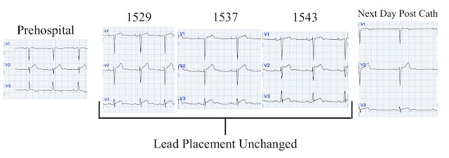
A prehospital EKG was also available for Patient 2:
This
EKG looks like the South Africa Flag Sign, indicating high lateral OMI.
It should be treated as such unless there is more information such as old or
serial EKGs that can confirm a benign diagnosis, as BTWI patterns can mimic the
South Africa Flag Sign (Compare this EKG to case 4 here: https://hqmeded-ecg.blogspot.
Patient 2, EKG 2 at 1537:
Marquette 12 SL algorithm read: ACUTE MI/STEMI
Patient 2, EKG 3 at 1543:
Marquette 12 SL algorithm read: ACUTE MI/STEMI
One examining these serial EKGs may note the diminishing S wave depth in V3 and become worried about terminal QRS distortion. V3 has a J wave that becomes more prominent as the S wave becomes smaller, however. This is not consistent with TQRSD which cannot have an S wave or a J wave in V2 and/or V3.
Still,
such dramatic changes cannot be overlooked. See this case, where a
patient with BTWI morphology and dramatic EKG changes within minutes is
diagnosed with myocarditis: https://hqmeded-ecg.blogspot.
EKG 3 also has a saddleback morphology in V2, which is only rarely due to OMI.
V2 has some features of type 2 Brugada phenocopy. The angle of
the downslope of the saddleback-shaped T wave (beta angle) is greater
than 35 degrees, consistent with type 2 Brugada morphology. Transient
Brugada morphology has been observed in patients with fever, on sodium
channel blocking drugs, or with hyperkalemia. The patient had none of
these conditions.
Whether these EKGs show myocarditis, a normal variant, or something else, they are overall not typical of transmural ischemia of the anterior or high lateral walls. Additionally, a bedside echocardiogram showed no wall motion abnormality and normal LV function.
The two cases were considered:
Patient 1 was recognized by the ED provider and the cardiologist as having resolved “STEMI”. He was given heparin and the decision was made to delay his catheterization until the next morning.
Patient 2 was diagnosed by the cardiologist with acute “STEMI” and he was taken emergently to the cath lab. Angiography revealed a 30% nonobstructive stenosis of the mid LAD. Serial high sensitivity troponin T (URL 15 ng/L) values were negative and stagnant.
Patient 1 remained in the hospital overnight. He had multiple episodes of bradycardia and nonsustained ventricular tachycardia. He went to the cath lab at 0900 the next morning. There was a 70% culprit stenosis of the first obtuse marginal branch in a right dominant system. It was stented. His high sensitivity troponin T trended from 69 ng/L on ED arrival to 583 ng/L two hours later, and peaked at 5,258 ng/L overnight. This was a large OMI. This is a good demonstration that when the artery reperfuses, it is at high risk of re-occlusion (in this case reoccluson/reperfusion/reocclusion/reperfusion).
A formal echocardiogram for patient 2 showed normal LV size, wall thickness, and global systolic function. He had another EKG taken the next morning:
The BTWI pattern is less prominent, but persists.
Patient 2 refused further workup and was discharged on day 2. The fluctuations of his BTWI pattern can be appreciated below:
A formal echocardiogram for patient 1 showed:
Moderately increased LV size
Reduced global systolic function with an estimated EF of 35-40%
Mid and distal anterior wall, mid and distal anterior septum, entire apex, and mid septum segment wall motion abnormalities. Much of this is due to his prior LAD and Cx OMI.
An EKG for patient 1 was taken after catheterization:
The lateral T waves show terminal inversion, consistent with reperfusion. There are well formed Q-waves in lateral precordial leads.
How did the Queen of Hearts perform?
Patient 1:
Prehospital EKG- OMI with high confidence
ED EKG- OMI with high confidence
Patient 2:
Prehospital EKG- OMI with high confidence
ED EKG 1- OMI with mid confidence
ED EKG 2- OMI with mid confidence
ED EKG 3- OMI with high confidence
A disappointing false positive by the Queen, but she did not miss the subtle OMI.
Click here to sign up for Queen of Hearts Access.
MY Comment, by KEN GRAUER, MD (10/15/2024):
- I limit my comments to assessment of the initial ECGs from these 2 patients that were recorded in the ED. For clarity in Figure-1 — I’ve labeled these 2 ED tracings.
- NOTE: Although clinical priorities in today’s case became clearer when prehospital ECGs were revealed — ED physicians must often make decisions without the benefit of prehospital tracings (that are not always immediately available to ED physicians). I therefore thought it worthwhile to review the decision-making process solely from the perspective of the initial ED tracings.
- The patient is a 75-year old man with known coronary disease, including prior LAD and LCx OMI.
- He presents with new-onset CP (Chest Pain) — brought on by activity, and persistent for 3 hours, being severe enough to prompt helicopter transport to the ED.
- The initial ED ECG for Patient #1 (TOP tracing in Figure-1) — shows sinus rhythm — low voltage in the limb leads — normal intervals and axis — and no chamber enlargement.
- My "eye" was immediately drawn to lead III (within the RED rectangle). Considering the tiny amplitude of the QRS complex in this lead — I thought the upright T wave in this lead to clearly be more voluminous than-it-should-be, which in this patient with new and now persistent CP, qualifies as a hyperacute T wave.
- To emphasize that we clearly see evidence of prior infarction in this initial ED ECG from Patient #1 — as there are large, wide Q waves in multiple leads (ie, leads II,III,aVF; V5,V6) — with loss of r wave from lead V1-to-V2 + fragmented QS complexes in V3,V4. That this patient has severe underlying coronary disease is indisputable.
- BUT, in addition to the hyperacute T wave in lead III — the T waves in the other inferior leads are equally hyperacute (RED arrows within the BLUE rectangles).
- Note that the tiny QRS complex in lead aVL manifests T wave inversion (BLUE arrow in this lead) — consistent with reciprocal ST depression in response to an acute inferior OMI.
- Acute changes were also seen in the chest leads of this initial ED ECG from Patient #1 (within the PURPLE rectangles). Thus, the shelf-like ST depression (PURPLE arrows in leads V2,V3) — and the hyperacute-looking ST elevation in lead V6 (within the dotted PURPLE oval) strongly suggest associated acute postero-lateral OMI.
- BOTTOM Line: Patient #1 demonstrates acute infero-postero-lateral OMI that is superimposed on severe underlying coronary disease with prior infarctions. Prompt cath with PCI is clearly needed for Patient #1 on the basis of this initial ED ECG.
- P.S.: Without a prior tracing for comparison — it's impossible to know if the low voltage in the limb leads is an acute finding. This is relevant — because among the causes of new low voltage is myocardial "stunning" from a large acute MI, such that this ECG finding may serve as a harbinger of a reduction in LV function that may soon be occuring (See My Comment at the bottom of the page in the November 12, 2020 post of Dr. Smith's ECG Blog).
-labeled-USE.png) |
| Figure-1: I've labeled the initial ED ECGs in today's case from Patient #1 and from Patient #2. (To improve visualization — I've digitized the original ECG using PMcardio). |
- This patient is a 58-year old black man. He had a history of substance abuse — but no prior history of coronary disease. His CP began shortly after eating.
- To emphasize that the history for Patient #2 in no way rules out the possibility of an acute cardiac event — but it does sound less worrisome to me.
- My "eye" was immediately drawn to lead V4 (within the RED rectangle). As per Dr. Smith — the shape of the QRST complex in lead V4 just "looks" like BTWI (Benign T Wave Inversion) — in that there is fairly tall R wave amplitude in a lateral chest lead, in which there is a J-point notch (PINK arrow) — slight ST elevation with terminal T wave inversion (RED arrow) — and a relatively short QTc interval (For more on BTWI — See the March 22, 2022 post, in which Dr. Meyers shows a series of BTWI cases "in all of its flavors" — with My Comment on BTWI at the bottom of the page).
- Elsewhere on this initial ED ECG from Patient #2 are the following: i) Slight ST elevation in lead aVL, with terminal "slurring" of the QRS complex that is consistent with a repolarization variant (PINK arrow in this lead); and, ii) Benign-appearing small, rounded positive T waves in lateral leads V5,V6 — also consistent with a repolarization variant.
- There is T wave inversion in lead III — but this is not necessarily abnormal given the RSr' complex in this lead.
- The principal finding of concern relates to the leads with the question marks. The ST-T wave in lead V2 looks larger than I would expect given modest size of the QRS complex in this lead. Of even more concern — is the J-point ST elevation with straightening of the ST segment takeoff in lead V3. In this context — I could not rule out potential significance of the slightly elevated and straightened ST segment in lead V1 — and, I felt a need to "relook" at the ST elevation in lead V4.
- BOTTOM Line: I did not feel comfortable ruling out the possibility of an acute cardiac event on the basis of this ED ECG from Patient #2. That said — I would not activate the cath lab on the sole basis of this tracing, but instead would get more history — repeat the ECG within 10-to-20 minutes — check serial Troponins — and do a bedside Echo during chest pain, looking for a wall motion abnormality.
- The prehospital ECG of Patient #1 — showed an obvious acute STEMI. The PEARL to remember — is that by correlating the initial ED ECG with this patient's total resolution of CP at the time this ED ECG was recorded — we can establish that spontaneous reperfusion had occurred. This clinical correlation of patient symptoms to the timing of each ECG also establishes the need for prompt cath with PCI to prevent reocclusion (ie, A "culprit" vessel that spontaneously reopens — may just as easily spontaneously reclose if not promptly treated with PCI).
- Patient #2 — had a series of insightful prehospital ECGs done, which showed marked change in ST-T wave appearance from one tracing to the next. As emphasized by Dr. Smith — the PEARL to remember is that "BTWI patterns can be very dynamic". As a result — great caution is needed so as not to misdiagnose the potential changing ECG pattern of a patient with BTWI as having dynamic ischemic changes. This can be tricky! — such that cardiac catheterization of Patient #2 in today's case was not at all inappropriate (albeit I thought Patient #1 merited priority for the cath room).


No comments:
Post a Comment
DEAR READER: I have loved receiving your comments, but I am no longer able to moderate them. Since the vast majority are SPAM, I need to moderate them all. Therefore, comments will rarely be published any more. So Sorry.