42 y.o. male with no past medical history presented for chest pain of onset 2 weeks prior. It is not constant, but lasts only a couple minutes. It is substernal without radiation, and is associated with SOB. Onset of chest pain was 2 weeks. States it is not constant.
A few days prior, his chest pain was intense and lasted about 10 minutes and it made him sweat.
On the day of presentation, he was walking to the ED from the parking lot and the chest pain recurred and lasted about 2 minutes.
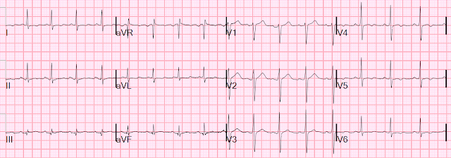
Here is his ED ECG:
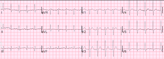
Here is a previous ECG:
The T-wave changes were not present at that time.The first high sensitivity troponin I returned at 32 ng/L. The 99th percentile upper reference limit (URL) is 34 ng/L for men, and 16 ng/L for women; since this patient is a man, this level does not quite qualify for acute MI.
4th Universal Definition of Myocardial Infarction requires:
1) a rise and/or fall in cTn with at least one value above the 99 the percentile occurring in appropriate clinical circumstances consistent with acute myocardial ischemia. AND
2) 1 or more of the following: a) ischemic symptoms, b) development of pathological Q waves in the 12-lead ECG, c) ECG changes indicative of new ischemia, d) imaging evidence of new loss of viable myocardium or new regional wall motion abnormality, or e) identification of an intracoronary thrombus by angiography or autopsy.)
Since this troponin is not above the 99th percentile URL, it is not acute MI (yet). But perhaps the next one will be higher and we can make the diagnosis of MI?
Educational interlude on troponins: Why is 32 ng/L normal, and not high?
The manufacturer determines normal from a population of asymptomatic "normal" people without known heart disease who are without symptoms. The value below which 99% of normals are measured is defined as the URL. For this Abbott Architect high sensitivity troponin I, that value is 16 ng/L for women and 34 ng/L for men. However, individuals with measurable values below the 99th percentile (below the URL but above the limit of detection - LoD) have >3x the incidence of adverse outcomes at 180 days compared to those whose values are reported as below the limit of detection (LoD), which is 1.9 ng/L (Europe). [U.S. FDA only allows reporting the LoD at 4 ng/L]. This date comes from our publication in Circulation.
Dr. Fred Apple and others studied many assays in individuals who are even more certain to be "normal." (Clinical Chemistry, Volume 66, Issue 3, March 2020, Pages 434–444) as proven by a normal GFR, normal Hemoglobin A1C, and normal BNP. In this asymptomatic group with no evidence of serious illness, the values for this Abbott Architect assay (he studied several other assays also) were 13 ng/L for women and 20 ng/L for men.
So for a very healthy patient, a value of 32 ng/L is actually high!! Nevertheless, strictly speaking, it cannot be used to make the diagnosis of acute MI. But it sure can make you suspicious that ACS is the etiology, and thus the diagnosis is Unstable Angina.
How about this patient? His A1C = 9.2, GFR is normal, and BNP is normal. It is uncertain what the 99th percentile for a group of patients with normal GFR, normal BNP, but elevated A1C would be.
Another ECG was recorded:
The 2nd troponin returned at 33 ng/L. So the "delta" is an insignificant 1 ng/L (this difference is within laboratory imprecision). Our data and our protocol would say that if both troponins are below the 99th percentile URL (34 ng/L) and the delta is < 3, then the patient would not rule in for MI if more troponins (e.g., at 6 or 12 hours) were to be measured. Thus he is eligible for discharge.
However, patients with a classic history, as here, must not be discharged, regardless of the ECG or troponin!
Let's do a HEART score: worrisome history, 4 risk factors, and nonspecific ECG = 5 (moderate risk)
How about EDACS score: = 15 (< 16 is low risk)
So the patient was admitted.
-------An alternative would be a CT Coronary Angiogram, but this is only available 8AM - 4 PM on weekdays. Also, since this patient's symptoms were provoked by exertion, a stress test is a very good test for him.
Next day he had a stress echo, which was positive and is interesting:
The patient exercised for 7:25 minutes on the standard Bruce protocol and achieved a peak heart rate of 153 bpm representing 86% of age predicted maximum heart rate and an estimated work load of 9.1 METs. Test was terminated because of chest pain and dyspnea.
Abnormal stress echocardiogram with a high degree of certainty--evidence for ischemia in the LAD territory.
1. Inducible wall motion abnormality with stress--apical septal, mid anteroseptal, mid to apical anterior, apical lateral, and apical inferior akinesis with stress.
2. Left ventricular function normal at rest, severely decreased with stress.
3. Left ventricular dilatation with stress.
4. Ischemic ECG response with stress (up to 2 mm of flat ST depression in the inferior leads, most severe 3:00 minutes into recovery).
5. The patient reported angina with stress.
6. Exercise capacity was fair (85% METS for age and gender).
7, Hypertensive response to exercise (peak BP 215/111 mmHg).
Angiogram:
Severe 3 vessel disease.
Required 5 vessel CABG.
Learning Points:
1. Unstable Angina still exists.
2. A troponin beneath the diagnostic threshold for myocardial infarction is not always normal.
3. A strong history should supercede a negative ECG and troponin.
- Just because a patient rules out for an acute event — in no way rules out the possibility that this patient might still have underlying coronary narrowing.
- The history that this previously healthy patient gave, was of chest pain that began 2 weeks earlier. The pain was not constant. It lasted only minutes (by his account — never more than 10 minutes). The pain was substernal, without radiation — and associated with shortness of breath. He did report one 10-minute episode of more severe chest pain associated with diaphoresis — but that was several days earlier. Other than on the day of admission — there was no mention in the history provided of exacerbating or alleviating factors.
- From the history given — we do not know WHY this patient chose the particular day that he walked into the ED. The only mention of chest pain on that day was walking from the parking lot, and that episode lasted only 2 minutes.
- NOTE: It is true that "deniers" often minimize (or completely deny) symptoms — but at least on paper — the history we are given did not sound to me like the history of an acute event. That said — of obvious concern in this 42-year old man with no prior history of chest pain, were his 4 significant coronary risk factors (ie, smoking, hypertension, hyperlipidemia and diabetes).
- KEY Point: The literature suggests that for a patient of a "certain age", who presents with new chest pain to the ED — that cardiac risk factors play a minor role in influencing the likelihood of whether the patient is having an acute cardiac event. In contrast — the likelihood of underlying coronary disease is greatly influenced by the presence of multiple cardiac risk factors (such as those described for today's patient).
- Editorial NOTE: In my experience — there is a unconscious "self-selection" that chest pain patients intuitively make at the time they decide whether to go to their family physician's office, to an Urgent Care Center — or to the ED. By this I mean, that given the identical history and risk factors — statistical likelihood that a patient is having an acute event is much greater for those patients who come to the ED, compared to those patients who decide to go to the office. In my 30 years of seeing ambulatory patients and precepting countless residents — it was exceedingly uncommon for a patient with an acute MI to present to our office. Instead, almost all of our patients who had acute MIs "self-selected" and went to the ED.
- As per Dr. Smith — although this patient's Troponin values of 32 ng/L and 33 ng/L were slightly below the acute MI "cut-off" — these values were clearly higher-than-expected when compared to a healthy population. I would not call these Troponin values "completely normal".
- That said — even IF this patient's serum Troponin values and his serial ECGs were totally normal — this in no way would rule out the possibility of underlying coronary disease. On the contrary, given this patient's 4 cardiac risk factors and his history of new symptoms — some form of Stress Testing is clearly indicated.
- IF instead of presenting to the ED, this patient had presented to our Primary Care Clinic — We would have followed the identical approach that was followed after ruling out an MI ( = Risk stratification by some form of Stress Testing).
- For clarity — I've reproduced the 3 ECGs from today's case in Figure-1. I've rearranged the sequence that I show these 3 tracings to facilitate comparison between this patient's initial ECG ( = ECG #1) — with his "baseline" tracing ( = ECG #2) — and then between his initial ECG with the repeat ECG done a little bit later in the ED ( = ECG #3).
-USE.png) |
| Figure-1: Comparison between the 3 ECGs in today's case. |
- ECG #2 ( = the "Baseline" tracing) — The rhythm is sinus tachycardia at the fairly rapid rate of ~115/minute! There is a Q wave of uncertain significance in lead III. There is no chamber enlargement. R wave progression is normal. There is some nonspecific ST-T wave flattening in a number of leads — but nothing that looks acute. KEY Point: Given the significant tachycardia — true comparison of ECG #2 with the 2 ECGs done the day the patient presented can not be accomplished until we understand the circumstances causing this tachycardia.
- ECG #1 ( = the initial ECG in Today's Case) — The rhythm is sinus. The heart rate varies (ie, sinus arrhythmia) — but overall, the rate is clearly slower than it was in ECG #2. There is no significant change in either frontal plane axis or precordial lead QRS appearance — so other than the change in heart rate, comparison of ST-T wave change between ECG #1 and ECG #2 should be valid. As noted earlier by Dr. Smith — there is now shallow T wave inversion in lateral chest leads V4,5,6 that was not present in ECG #2.
- ECG #3 ( = the tracing done after ECG #1 in the ED) — A regular sinus rhythm at ~80-85/minute is seen (the rate slightly slower than for ECG #1). There has been a change in frontal plane axis (ie, the QRS is now predominantly negative in lead III — whereas it was positive in ECG #1). This may make it more difficult to evaluate any "true" change in limb lead ST-T wave morphology. I thought there was a slight-but-real change in ST-T wave appearance in the chest leads (ie, the T waves in V1,V2 are smaller — and there was a bit more T wave inversion in other leads).
- Acute OMI has essentially been ruled out.
- This 42-year old man with new symptoms + 4 cardiac risk factors + 2 serum Troponins that are at least suspicious + subtle but-I-think-real ECG changes — is clearly in need of Stress Testing.
- The abnormal Stress Echo — especially in view of development of chest pain with exercise in association with both Echo and ECG evidence of ischemia — dramatically increases the post-test likelihood of significant coronary disease. I would have been surprised if significant coronary was not found on cath.
- I strongly suspect the 42-year old man in today's case was a "minimizer" of symptoms. I would not be at all surprised if after doing the Stress Test — a longer duration and much more typical history for anginal symptoms could have been obtained.


No comments:
Post a Comment
DEAR READER: I have loved receiving your comments, but I am no longer able to moderate them. Since the vast majority are SPAM, I need to moderate them all. Therefore, comments will rarely be published any more. So Sorry.