Monday, November 29, 2021

A Pathognomonic ECG. What is it? (Hint: 2 diagnoses in one)

I was reading through the list of EKGs and saw this one.  What is it diagnostic of? (hint: 2 diagnoses in one)


There is a very long, flat, ST segment, resulting in a long QT (most long QT is due to a wide T-wave, not a long ST segment).  This is diagnostic of hypocalcemia.  There are also peaked T-waves of hyperkalemia.  This is a common combination in dialysis patients.

The ionized calcium was 2.29 mg/dL (normal is 4.40-5.20).  The K was 6.2 mEq/L.

Here are the symptoms she had (very typical for hypocalcemia):

Dialysis patient with left upper extremity numbness and tingling, lightheadedness, perioral numbness and tingling, and bilateral lower extremity stocking glove numbness and tingling. 

She was given 6 grams of calcium gluconate and K was shifted.

Here is the subsequent ECG:

All findings are corrected


Saturday, November 27, 2021

A man in his 40s with epigastric pain and a dynamic ECG

 Case written and submitted by Dr. Arjun J V, peer reviewed by Meyers, Smith, Grauer


A 49 year male patient was brought to our ED at around 9 PM on with complaints of epigastric pain since that afternoon. The patient had the same complaints on and off for many years which would resolve on taking OTC antacids. However, this time the pain was persistent and included new diaphoresis, so he presented to an outside facility where this ECG was recorded:


What do you think?


There is sinus rhythm with a narrow QRS complex with normal axis. There is slight PR depression in III, followed by some STE with upright T wave. Lead aVL shows a negative QRS complex with a seemingly large negative T wave. Lead I also has a negative T wave. 

There is STE in the anterior leads but with reassuring large voltage QRS and reassuring morphology; to me it fits with normal variant anterior STE, not concerning for OMI. 

The question is: is this concerning for subtle inferior OMI? On on hand, the T wave in III is not definitively hyperacute, but the T wave in aVL may be fairly big for its QRS complex. We have shown many examples of this. 


At the outside hospital, the patient was given DAPT and was referred to a higher centre with Cath lab facilities. The providers were concerned for inferior OMI. The patient was hemodynamically stable but had persistent discomfort. An ECG was obtained at the receiving facility on arrival: 


What has changed compared to the first ECG? What does it mean?


Now the STE and T wave in III seem to be less elevated than before. The deep TWI in I and aVL are replaced by upright T waves. In the first ECG, aVR has a slightly upright T wave (a red flag for this condition, by the way), whereas now it is depressed.

Does it mean that the inferior OMI is starting to reperfuse? Does it mean that the high lateral area is showing pseudonormalization??

NO!

Neither of these are correct. All changes between the two ECGs are explained completely by reversal of the LA and RA electrodes on the first ECG.


Look at the differences:

 - Lead I (defined by RA negative, LA positive) is perfectly inverted between the two ECGs: in the first ECG (with lead reversal) we see qR then negative T wave. In the second ECG (with correct leads) we see rS with positive T.

 - Lead "aVL" in the first ECG is actually lead aVR, and it matches lead aVR of the second (correct leads) ECG

 - Lead "aVR" in the first ECG is actually lead aVL, and it matches aVL in the second (correct leads) ECG

 - aVR should not generally have a positive T wave if the leads are correct and the QRS is narrow and overall leftward


Below is Ken Grauer's excellent diagram showing the effects of LA/RA reversal:


Here they are side by side:


Here is the initial ECG before and after correcting the leads as per Dr. Grauer's diagram:


Unfortunately the lead reversal was not noticed by the treating team, who was still worried about OMI.

The patient’s ROS was unremarkable and POCUS did not show any RWMA. Both the ED team and Cardiology team were not completely convinced for the need for an emergent CAG but erred on the side of caution, possibly because of the following reasons:

 - Initial ECG showed perceived changes of inferior wall MI 

 - Young MIs can present with atypical changes

 - The recent & sudden death of a 45 year old male celebrity due to MI who was known to be extremely fit and health conscious. The untimely death shook the whole state to the core which witnessed an increase in the number of patients getting health check-ups[1]


Troponins were not awaited and emergent angiogram was done which was completely normal, with no evidence of CAD. No complications of the angiogram were experienced.


Labs were unremarkable. Troponin-I, Lipase & Amylase were negative.


CT abdomen showed possible changes of acute calculous cholecystitis. Patient was advised surgery but the patient did not go through with it. Patient was discharged against medical advice. Further follow up is unavailable.


Learning Points:

Lead reversal is common and can create scenarios like this one, in which a well meaning physician looking for signs of OMI can be fooled into concern for a dynamic ECG finding, leading to unnecessary concern and possibly an unnecessary angiogram. Learning the most common forms of lead reversal can help prevent this.

LA/RA reversal causes lead I to become inverted, and switches the places of leads II/III and aVL/aVR, while lead aVF remains unchanged.

When the QRS and T waves are otherwise normal, a positive QRS and/or T wave in aVR can be a red flag for lead misplacement.

Human factors and recency bias can affect patient management.


References:

1. https://bangaloremirror.indiatimes.com/bangalore/others/a-not-so-hearty-tale/articleshow/87475651.cms


===================================

MY Comment by KEN GRAUER, MD (11/27/2021):

===================================

Lead misplacement is easy to overlook! This is because of the tendency to "assume" that routine normal placement of extremity electrode leads will automatically happen. And because it almost always does happen — we are not used to recognizing lead misplacement when it does occur. There are several additional reasons why the LA-RA lead reversal may have been missed in today's case:

  • This patient has RAD (Right Axis Deviation) on his baseline tracing — with a small amplitude QRS complex showing predominant negativity in lead I. LA-RA lead reversal is usually picked up because of the finding of "global negativity" (of the P wave, QRS and T wave) in lead I. But because of the RAD — the QRS complex in today's initial tracing was predominantly positive! (and therefore simulated a high lateral Q wave infarction with deep T wave inversion).
  • Global negativity (ie, negative P wave, QRS and T wave) is normally seen in lead aVR — because this most remote electrode lead normally views the heart's electrical activity as traveling away from its distant perspective (looking down from the right shoulder). But because of the baseline RAD — we did not see an all upright R wave as a "tip-off" to LA-RA lead reversal in lead aVR.
  • Small amplitude P wave activity in lead I on the initial tracing in today's case did not clearly suggest a negative P wave in lead I, that also would have been a "tip-off" to LA-RA lead reversal.
  • Finally — the P wave in lead II was upright, so that sinus rhythm was assumed.


The diagram Dr. Meyers credited me above, showing the effects of LA-RA lead reversal on the ECG is adapted from the LITFL ( = Life-In-The-Fast-Lane) web site — which is my "Quick GO-TO" reference for the most common types of lead misplacement. Simply put in, "LITFL lead reversal" in your on-line Search bar — and this KEY link comes up instantly!

  • LA-RA lead reversal is by far the most common technical mishap. It is usually EASY to recognize, because we are likely to see: i) Global negativity in lead I — which should never normally be seen — and which tells you there is either lead misplacement or dextrocardia; ii) The QRS in lead aVR is upright; andiii) The P wave in lead II will often be negative.
  • For another example of LA-RA Lead Reversal with more detailed discussion of this entity — CLICK HERE.


In Summary:

There are several reasons the LA-RA lead reversal was easy to overlook in today's case. The reason I immediately picked it up — is that today's initial ECG simply "looked funny" to me — because:

  • It is highly unusual to see a very wide Q wave with such disproportionately large T wave inversion in lead I (even when you have acute high lateral infarction).
  • We do not see global negativity in lead aVR (ie, the P wave is flat and the T wave is definitely positive).
  • Although positive — the P wave in lead II is smaller than the P wave in lead III (which would be unusual for normal sinus rhythm).
  • Lead aVL "looks funny" (ie, it shows "global negativity" — that is usually seen in lead aVR).
  • KEY Point: Whenever I see an ECG that "looks funny" — I verify lead placement and then immediately repeat that tracing (especially when my differential diagnosis is "Rule Out" acute MI)!


Thursday, November 25, 2021

Any ST depression in V2 and V3 is posterior OMI until proven otherwise, especially if downsloping

A middle aged male presented after onset, approximately 50 minutes prior, of constant crushing 10/10 substernal chest pain, radiating into right arm associated with shortness of breath. He had never felt this way before. There was a history of HTN but he was not taking any medicines.

Prehospital ECG was recorded approximately 20 minutes after pain onset and 20 minutes prior to ED arrival:

There are somewhat large T-waves in II and aVF and a sagging ST segment in aVL, suggestive of inferior OMI.  
There is some minimal downsloping ST depression in V2 and V3, which is suggestive of posterior OMI. 

The ECG is not diagnostic however.


This first ED ECG was recorded approximately 10 minutes after ED arrival, and about 30 minutes after the prehospital ECG:

There is downsloping minimal STD in V2 and V3.  
The T-wave in lead II is somewhat suspicious, but is not a diagnostic OMI finding.
This is highly suspicious for isolated posterior OMI


Here are the precordial leads magnified, and the ST segments in V2 and V3 highlighted

There is perhaps a 0.5 mm of ST depression in leads V2 and V3, and it is downsloping.


In our recent research, published in November 2021 in The Journal of the American Heart Association, any ST depression maximal in V1-V4 is 96% specific for OMI requiring PCI (assuming, of course the patient has symptoms compatible with ACS).  

It is important to remember that the ECG in normal people without ischemia and without an abnormal QRS such as RBBB never has more than 0.5 mm of STD, and rarely has any STD at all.  The vast majority have some STE in V2 and V3.  (Macfarlane PW. Age, sex, and the ST amplitude in health and disease. J Electrocardiol [Internet] 2001;34 Suppl:235–41.)

In our study: There were 118 patients with "suspected ischemic ST depression maximal in V1-V4" (45 with less than 1 mm).  I blindly read ECGs of all 808 patients, and called OMI in 112 of these 118 patients; 99 had OMI and, of these, 34 had less than 1 mm STD.  Of these 34, 12 had STEMI in other leads, but 22 did not.  Of these 22, every one had some other subtle OMI findings in other leads.  In other words, we did not have a single patient with STD maxV1-4, AND less than 1 mm who did not have some subtle finding elsewhere, as is the case here On the other hand, we did have 2 patients with less than 0.5 mm STD and no other OMI findings whom I interpreted as "no OMI" who indeed had no OMI.  

So the finding of less than 1 mm in V2 and V3 which is completely isolated (no OMI findings elsewhere on the 12-lead) remains highly suspicious, but not diagnostic.  

However, downsloping STD in V2 and V3 is, in my experience, diagnostic.  

Case continued

I was very worried about about OMI from the crushing chest pain alone, even without the ECG, and I was specifically worried about posterior OMI for the above reasons. 

6 minutes later, we recorded posterior leads:

This shows a trace of STE in V4, but not up to 0.5 mm.  
I think the STD in V2 and V3 may have deepened a bit, but I did not notice it at the time.

Case continued

The blood pressure was 175/125.  This made aortic dissection a possibility as well.  We started him on high dose IV nitroglycerin up to 200 mcg/min, with sublingual NTG every 5 minutes, and this brought his BP down to 120/73. The pain persisted.  This is important: ACS with pain that can't be controlled with NTG should go emergently to the cath lab "regardless of ECG or biomarker findings!" (Eur Soc of Cardiology guidelines)

I called the cardiologist directly and stated that I thought this was ACS/OMI.  He agreed and we decided to do a CT for dissection.  If that was negative, the patient would go emergently to the cath lab.

While I was on the phone with him, we recorded this ECG 17 minutes after the 2nd one (23 minutes after the first), with posterior leads still attached:

This one has evolved and is now definitely diagnostic.  
Increased STD in V2 and V3
New STE in I and aVL
New hyperacute T-wave in I
Increased T-wave size in aVL
New inferior reciprocal ST depression.  
Greater than 0.5 mm of STE in posterior leads V8 and V9 (labelled V5 and V6)


The cath lab was activated.


While waiting for the cath team, another ECG was recorded at 37 minutes after the first, standard 12 lead (not posterior):

Now there are additionally hyperacute T waves in V4-V6, with some new ST elevation


The initial troponin I returned at 10 ng/L, which is within the normal range.

Aside: In our study of 925 patients with STEMI, using the same assay, the initial troponin was below the 99th percentile in 14.4%  Here is the reference: Wereski et al. (including Smith).  High-Sensitivity Cardiac Troponin Concentrations at Presentation in Patients With ST-Segment Elevation Myocardial Infarction

Angiogram and PCI

1. Acute coronary syndrome.

2. Thrombotic D1 occlusion 100% (this is the culprit vessel).

3. Multivessel coronary artery disease 

4. Successful D1 stenting with a drug eluting stent.


Post PCI


Next morning


The troponin I peaked at greater than 50,000 ng/L, a very large acute OMI.


Echo:

Mildly increased LV wall thickness with normal LV cavity size and an estimated ejection fraction of 45%.

Regional wall motion abnormality--mid anterolateral and mid inferolateral hypokinesis.


This is consistent with posterior OMI ("posterior" is now classified as "lateral" -- this is complicated and explained in the article).


Learning points:


1. Any ST depression that is not explained by RBBB or other QRS findings, in a patient with symptoms suggestive of ACS, is posterior OMI until proven otherwise.

2. Unexplained chest pain that is typical of ACS, and is not controlled with medical therapy, needs an angiogram even in the absence of ECG or biomarker proof of ACS.

3.  ST depression of posterior OMI is almost always accompanied by some other subtle OMI finding in inferior or lateral leads.

4. ST depression that is downsloping, even if less than 0.5-1.0 mm, is particularly worrisome, even in the absence of other subtle OMI findings.

5. When there is severe hypertension, all chest pain might be due to supply demand mismatch.  Therefore, one must lower the BP before activating the cath lab.


Monday, November 22, 2021

Shark fin post arrest: do you understand the ECG?

Case submitted by Dr. Daryl Williams, written by Pendell Meyers, peer reviewed by Smith and Bracey


A physician bystander witnessed a middle-aged or slightly elderly man suddenly collapse while walking down the street, very close to the hospital. The physician immediately started CPR and called EMS. EMS arrived quickly and found the patient to be in VFib. After several shocks the patient achieved ROSC.

A minute or so after arrival to the ED, he went back into VFib and was immediately shocked back out into sinus rhythm.

His EMS ECG during initial ROSC was available for the ED team:


Here is his ED ECG:

What do you think?


Both ECGs above show RBBB + LAFB, with massive concordant STE in leads V2-V6 as well as I and aVL. There is shark fin morphology (aka "Giant R wave", or "Lambda wave"), in which the wide complex QRS appears to fuse with the massive STE, causing confusion as to where the J point actually is, and where the QRS ends and the ST segment begins. This pattern is diagnostic of at least LAD occlusion (which of course supplies the anterolateral walls and the RBB and LAF), but given how severe the findings are even out into the high lateral wall, the occlusion may even be more proximal, such as the left main. This pattern is more commonly seen in LAD occlusion simply because persistent left main occlusion is rare, probably because it such patients rarely survive long enough to obtain an ECG and/or angiogram.

Below, I have placed red vertical lines at the J points.

I was sent the above ECG with no clinical information, and my response was: "Should be a huge LAD or theoretically even left main occlusion."


The cath lab was activated immediately. At this time the patient had stable ROSC, was intubated, and did not require pressors.

The LV EF was estimated at 20%. An Impella was placed for hemodynamic support. 

Angiogram:

Acute thrombotic culprit lesion of the distal left main coronary artery and proximal LAD with 80% stenosis, also at the time of angiogram. There was also 80-90% stenosis of the proximal D1, and 90% at the ramus intermedius. It is not completely clear whether these were also thrombotic extensions of the distal left main lesion, but these are probably all part of the same acute culprit left main lesion, or downstream showering from it. 

There was mild diffuse (non-quantified) CAD of the LCX, otherwise no evidence of CAD in any other part of the coronaries.


As predicted from the ability to obtain ROSC, there is not full occlusion at the time of cath. There is flow in each vessel distally. 

For a patient who suffers cardiac arrest with recurrent or refractory VF due to left main occlusion, it is very unlikely to get stable ROSC unless the lesion opens up at least a little bit. And that is what we see here. The post cath ECG below confirms reperfusion including resolution of RBBB and LAFB:

In fact, now there is more of a LBBB pattern, but a bit odd because I and aVL are not all upright.

Initial contemporary troponin T was 0.11 ng/dL (reference range less than 0.01 ng/dL). Troponin rose to 2.10 ng/dL but then no further measurements were recorded, so the peak troponin was not measured.

He had a long and rocky course in the ICU. Brain MRI showed multiple areas of restricted diffuse and acute cerebral infarct consistent with hypoxic encephalopathy. Despite ideal cardiac arrest conditions (immediate professional CPR, near the hospital, quick response time, seemingly zero no-flow time, and aggressive immediate cath lab activation), it seems that there will be at least some element of permanent neurologic injury, but the severity is yet to be determined.


Learning Points:

You must understand the shark fin morphology, and the fact that the LAD supplies the right bundle branch and the left anterior fascicle, if you want to be able to diagnose the most severe OMIs that exist like this one! STEMI criteria do not apply here, and many providers will not even know where to measure the ST segment at all.

In this case, we see lead aVR has ST and T wave depression in lead aVR, NOT ST elevation. This is because aVR simply shows the reciprocal findings of the many other leads which are oriented in the opposite direction. With widespread STE in many leftward leads, it is inevitable that lead aVR must show reciprocal STD. If there had been better coronary perfusion and more collateral circulation at the time of these ECGs, then instead of an anterolateral OMI pattern, we may have seen the pattern of diffuse subendocardial ischemia, with diffuse STD and reciprocal STE in aVR. The key is that aVR has no new or primary information. I teach that "aVR" stands for the "aVerage Reciprocal" lead, because it shows the reciprocal findings of the average of all the other leads on the 12-lead, which are largely oriented in the opposite general direction from lead aVR.

Sometimes even with the best possible conditions, the neurologic outcome of cardiac arrest can eclipse the cardiac salvageability.

Some of the most severe LAD or left main occlusions present with acute RBBB and LAFB, and these findings carry the highest risk for acute ventricular fibrillation, acute cardiogenic shock, and highest in-hospital mortality when studied by Widimsky et al. (in-hospital mortality was 18.8% for AMI with new RBBB alone). Additionally, the RBBB and LAFB make the recognition of the J-point and STE more difficult and more likely to be misinterpreted (when the QRS is wide, the J-point will hide!). Upon successful and timely reperfusion, the patient may regain function of the previously ischemic or stunned fascicles.


Widimsky PW, Rohác F, Stásek J, et al. Primary angioplasty in acute myocardial infarction with right bundle branch block: should new onset right bundle branch block be added to future guidelines as an indication for reperfusion therapy? Eur Heart J. 2012;33(1):86–95. 

https://pubmed.ncbi.nlm.nih.gov/21890488/


See these other related posts:

A deadly alcohol binge: a man in his 30s with chest pain and initial high sensitivity troponin I within normal limits


See 7 more cases of true total Left main occlusion here:

Here is a post on RBBB + LAFB and its significance, with links to many more such cases:


See other "shark fin" morphology cases here:

Fascinating case of dynamic shark fin morphology - what is going on?


But don't be fooled by the other etiologies of shark fin morphology:

Another Shark Fin. With a twist.


Friday, November 19, 2021

A woman in her 60s with syncope and vomiting. Does she need a pacemaker?

 Written by Pendell Meyers with some edits by Steve Smith


A woman in her 60s on chemotherapy presented to the Emergency Department for a syncopal episode just prior to arrival. She was walking to the bathroom when she suddenly felt nauseous and passed out. EMS was called by the patient's daughter, and en route to the ED she vomited twice. On arrival to the ED, she adamantly denies chest pain but says she's "just still not feeling well." She had no prior known cardiac disease.

Triage at 0755:


The rhythm is most either atrial fibrillation with complete heart block and resulting junctional escape, or atrial flutter with very high degree (but constant) block. Remember, atrial fibrillation cannot result in regular ventricular rhythm if it is conducted through the AV node.

The QRS has narrow normal morphology. There is almost 1 mm STE in III, but no STE at all in II or aVF. The T wave in III is hyperacute, and is biphasic up-down. Leads aVL and I show reciprocal findings (STD and TWI biphasic down-up). Through the baseline wander, you can see the impression of downsloping STD in V2 indicating posterior extension. Interestingly, V3 then appears to have a hyperacute biphasic down-up T wave, with hyperacute Ts in V3 and V4, but then V5 and V6 do not.  All the biphasic T-waves suggest that there is some early spontaneous reperfusion happening.

Overall, it is diagnostic of inferoposterior OMI, likely RCA occlusion, likely also explaining the acute bradydysrhythmia.


The rhythm was noticed as pathologic, but none of the ischemic findings discussed above were initially noticed. It seems as though the providers felt that the problem was a primary bradydysrhythmia.

Initial troponin returned at 520 ng/L, and another ECG is recorded at that time (0900):


Findings are very similar to the first ECG. There is ongoing active transmural (full thickness) infarction. Still no STEMI criteria.


Anther ECG was done at 1000 (reason unknown):

This ECG shows some interval reperfusion compared to the prior ones, including slight deflation of the hyperacute T waves and terminal T wave inversion in III, and the opposite in V2.

At this point the patient was persistently hypotensive and bradycardic requiring epinephrine drip to maintain heart rate above 50 bpm. She was described as uncomfortable and short of breath. She continued to deny chest pain. 


The ED team consulted cardiology for symptomatic bradycardia, asking whether the patient should go for an emergent pacemaker. The cardiology team evaluated the patient, reviewed all the ECGs above, and apparently found no concern for ischemia and agreed that the primary problem was just the bradydysrhythmia.

They were in the process of consenting the patient for a pacemaker when I contacted the ED provider in real time (I happened to be actively reading ECGs for the department from home at that time). I explained my concern for RCA occlusion as the cause of her OMI findings on ECG, and as the cause for her bradydysrhythmia (because the RCA almost always supplies the AV node).

The ED team performed a bedside echo which showed an inferior wall motion abnormality.

The repeat troponin I was 595 ng/L.

Another ECG was performed at 1055:


The end of the T wave in III is difficult to see through the atrial waves. There are some features of reperfusion, but also some leads that seem to show ongoing ischemia.

I was told the patient was still persistently symptomatic despite some features of reperfusion on ECG. She still required epinephrine and norepinephrine drip and was short of breath.

The ED provider was  able to convince the skeptical cardiologist to perform an angiogram before placing a pacemaker. 

The angiogram showed an acute thrombotic proximal RCA occlusion (TIMI 0 flow). After PCI, there was 0% residual stenosis with TIMI 3 flow.  

Comment: TIMI-0 flow does not rule out some degree of microvascular reperfusion; collateral flow may account for the minimal ECG features of reperfusion.  In fact, in Wellens' studies which established Wellens' syndrome, in all cases there was perfusion, but 20% of these cases were in spite of an occluded artery, but through collateral circulation.) This is precisely the reason why we conceptually define OMI as: "Acute coronary occlusion or near-occlusion with insufficient collateral circulation, resulting in imminent full-thickness myocardial infarction."

The first ECG after cath was performed hours later, in the cardiac ICU:


Sinus rhythm with PACs. There is resolution of the STE and hyperacute T waves, as well as terminal T wave inversion in III. This confirms reperfusion. Not to mention the dramatic improvement in AV node function.

The ICU staff noted that, after PCI, her heart rate rapidly improved and she never required even a temporary pacemaker. Her epinephrine and norepinephrine requirement resolved within hours of PCI. 

In fact, within 24 hours the patient required metoprolol and amiodarone for rate control of intermittent atrial fibrillation with rapid ventricular response:


Later she was back in sinus rhythm:


She had several minor complications unrelated to ACS during her stay, but was ultimately discharged on day 5.


Learning Points:

The most important, deadly, reversible causes of bradycardia include acute RCA occlusion (OMI), hyperkalemia, and toxicity from beta blockers, calcium channel blockers, or digoxin. We in the ED must be the experts at recognizing these conditions on the ECG. 

If this patient had instead received a pacemaker rather than RCA reperfusion, her ECG would have shown a ventricular paced rhythm. Providers who cannot recognize OMI in normal QRS conduction may be even less likely to recognize it in ventricular paced rhythm, although we have shown that it can be reliably done with proper training: 

New Review: Diagnosis of Occlusion Myocardial Infarction in Patients with Left Bundle Branch Block and Paced Rhythms


Until we get AI that can learn ECG patterns, we might need human ECG experts to act like radiologists, so that EM physicians and cardiologists can have access to expert interpretation.

You must learn to recognize hyperacute T waves. 

This patient is STEMI(-) OMI that clearly benefits from emergent reperfusion.

Wednesday, November 17, 2021

Toothache, incidental Wide Complex Tachycardia

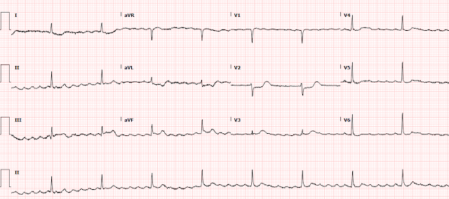
I was recently reading through the list of ECGs, and came across this one:

What do you think?


This was the interpretation I put into the system: 

"Re-entrant wide complex tachycardia with retrograde P waves, slow onset of QRS is c/w either antidromic WPW (ARVT) with pathway insertion in the superior RV, or with VT from the superior RV, most likely RVOT" (Right Ventricular Outflow Tract VT, one of the idiopathic VTs.


Explanation:

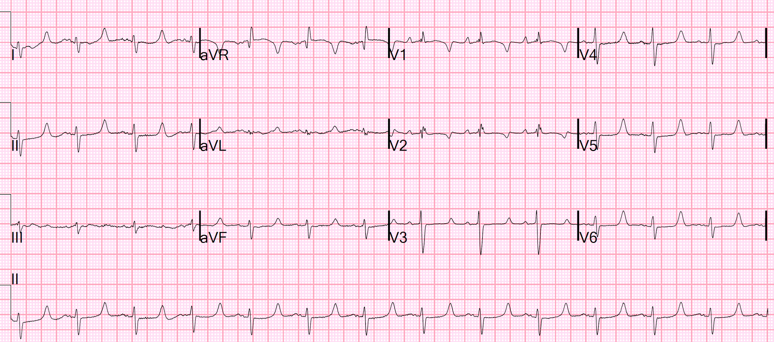
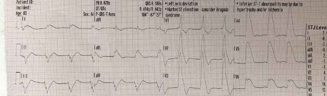
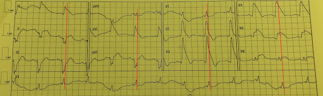
There is a regular wide complex tachycardia at a rate of 138.  Here are the important features:

1. V1 and V2 have an LBBB configuration (minimal or no R-wave, deep S-wave)

2. V5 and V6 have an LBBB configuration (monophasic R-wave)

3. Inferior axis (all upright in II, III, aVF)

4. The onset of the QRS is VERY slow (see slow downstroke in V1, V2, slow upstroke in other leads).  

    This slow onset means that it is either VT or AVRT (Atrioventricular reciprocating tachycardia).  AVRT is due to an accessory pathway (similar to WPW, but technically only accessory pathways with baseline delta waves can be called WPW), and when wide it is due to antidromic conduction (down the pathway resulting in pre-excitation of the ventricle -- delta wave with wide QRS).  It is the onset of the QRS that is slow -- NOT the latter part. 

If this is VT, it is coming from the RV (as there is LBBB pattern).  AND it is coming from the superior part of the RV so that the impulse is directed inferiorly (inferior axis).  This is typical of Right Ventricular Outflow Tract VT.   Other VT that comes from the RV and has LBBB pattern includes ARVD (Arrhythmogenic Right Ventricular Dysplasia, or RV Cardiomyopathy).   The latter does not necessarily have an inferior axis.

Apparently, RVOT is also called "Repetitive Monomorphic VT" as it comes in bursts or salvos.  At the ver bottom of the post, I have pasted part of an article from UpToDate, my favorite online resource, on this topic.

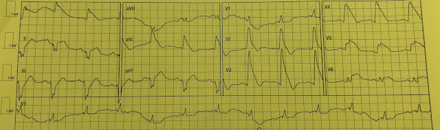
Here I annotate the ECG to show the retrograde P-waves

Arrows show the middle, or nadir, of the inverted P-wave.  
The inverted P-wave is not the upright component of the wave, but the trough.  
The blue line is drawn at the onset of the inverted P-waves

Case continued

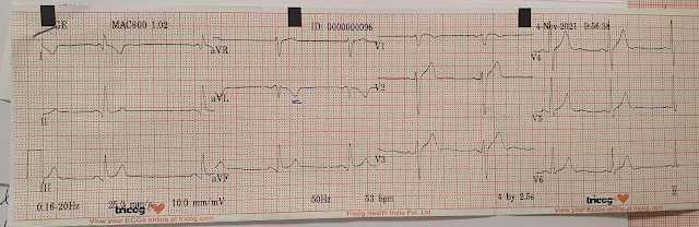
I looked to find the patient; she was still in the department and this is the story:

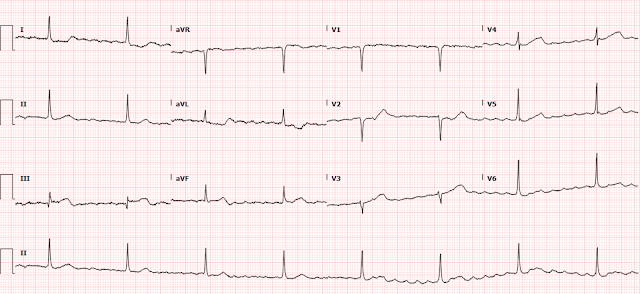
A 60-something woman with no cardiac history presented for tooth abscess.  Her pulse at triage was 76.  The intern elicited a history of chest pain 3 days prior and so ordered an ECG.  The intern was unaware of the heart rate at the time of the ECG until it was recorded:

Electrolytes including K and Mg were normal.

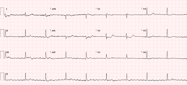
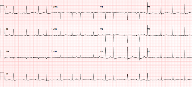
The patient spontaneously converted to sinus rhythm:

Now we see sinus tachycardia with PVCs and otherwise not too remarkable.  

What do you think?


My thought on this was that the PVCs have EXACTLY the same morphology as the wide complex tachycardia.  This indicates that the irritable focus of the tachycardia is in the ventricle at the exact point of the re-entrant rhythm, and therefore that the first ECG represents VT and not AVRT.  The absence of delta waves by itself is supportive but by no means diagnostic, as accessory pathways frequently do not manifest delta waves on the baseline ECG.  This is called "concealed conduction."  

See comments below by our electrophysiologist on the significance of the PVCs

Again, one does not call it WPW when there is concealed conduction; "WPW" is reserved for those with delta waves.  Nevertheless, whether WPW or not, accessory pathways offer another route for AV or VA conduction.

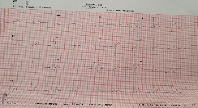
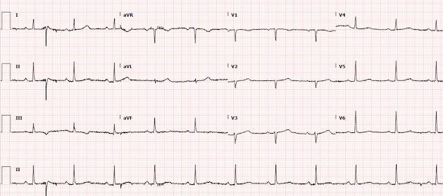
Shortly thereafter, she went back into tachycardia.  She was asymptomatic but was hypotensive at 80/50.


Same as before.  And it is not spontaneously converting

What do you want to do?


This is almost certainly RVOT, right ventricular outflow tract VT, and this kind of VT is responsive to adenosine!!  So if we could trust that she would not revert to VT, adenosine could work.  Alas, no short acting treatment such as adenosine or electricity will accomplish the goal of keeping the patient in sinus rhythm.  

Repeat: neither electricity nor adenosine will work to convert the rhythm.  Neither will solve the problem since we know that she can revert from sinus back to VT, as she has already done it once.  

Therefore, a long acting medication or an infusion (or both) are necessary.  Given the results of the Procamio Study comparing Procainamide to Amiodarone for stable monomorphic VT (for procainamide, it showed double the efficacy and half the adverse effects), we decided to give procainamide bolus (10 mg/kg over 20 minutes, with the option to give an additional dose of up to 7 mg/kg if necessary) and infusion.

2 g of Mg were also given.

After infusion of only 100 mg, she converted again. She remained in sinus rhythm on the infusion.
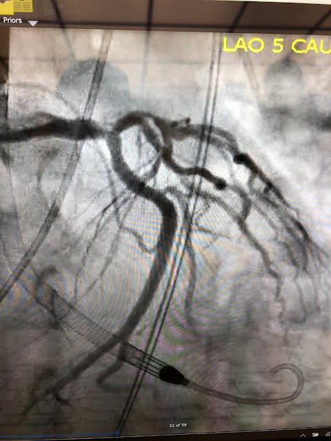


Echo:

--Frequent ectopy throughout exam.

--Normal LV size and wall thickness.

--Lower limits of normal left ventricular systolic function with an EF of 52%.

--There is no left ventricular wall motion abnormality identified.

--Normal right ventricular size and function.

--The estimated pulmonary artery systolic pressure is 34 mmHg + RA pressure.

--Based on the appearance of the IVC, the estimated RA pressure is normal.


Outcome:

The patient refused EP study, but the electrophysiologist was certain that this was Right Ventricular Outflow Tract VT (RVOT VT).  They discharged her on Diltiazem, which apparently works very well to prevent recurrent bouts of RVOT VT (I was unaware of this and could find no literature anywhere I looked).

Read more here:  Idiopathic Ventricular Tachycardias for the EM Physician 


Discussion by our Electrophysiologist

Smith: “I thought that the wide complex tachy (WCT) could be AVRT or VT”


EP: "Antidromic AVRT morphology would essentially be the same as “VT” originating from ventricular the insertion site of the accessory pathway. Therefore, traditional criteria for SVT with aberrancy do not apply to antidromic AVRT (except, that negative concordance can never be AVRT!)"


Smith: “But then when the patient converted and had PVCs of exactly the same morphology as the WCT, that it must be VT and not AVRT”


EP: "In cases of intermittent pre-excitation, you could potentially see wide and narrow QRS complex on the same EKG – but those of course will be preceded by P-wave (albeit with short PR interval)"


Smith: “Is this logic supported by evidence? Or is it still likely to be ARVT?”


EP: "Not sure, but PVC’s from RVOT and the wide complex tachycardia of same morphology is highly suggestive of it to be VT.

Another observation on the EKG is that presumably the retrograde atrial activity is quite far out from the next wide complex beat. In other words, if this was AVRT, then you’re assuming that the “PR” interval is very long and is conducting down the accessory pathway. Although there are slowly conducting accessory pathways, I believe they tend to cause Orthodromic AVRT more often. One example is “PJRT” that you might have come across.


On occasion, if someone has “fully pre-excited” beat (i.e., no fusion with AV node), then might be difficult to differentiate from a PVC as “P” wave might just be too close to the onset of QRS – but I don’t recall coming across such situation."


Finally:

In general, if you think a wide complex tachycardia is antidromic AVRT, then it’s “okay” to give AV nodal agent as AV node is still participating in the circuit. That’s whay you might have heard some folks mention using adenosine as diagnostic maneuver even in wide complex tachycardia if patient is hemodynamically stable (if it doesn’t terminate tachycardia, then might elicit AV dissociation if it is VT!). it does cause some vasodilatation, so I wouldn’t do it if someone already is hypotensive.


REPETITIVE MONOMORPHIC VT from UpToDateRepetitive monomorphic VT (RMVT) is characterized by frequent short "salvos" of monomorphic nonsustained VT (). It was first described by Gallavardin in 1922 and is variously described in the literature as RMVT, RV tachycardia, RVOT tachycardia, catecholamine-sensitive VT, adenosine-sensitive VT, and exercise-induced VT. Approximately 10 to 15 percent of cases arise from the left ventricular outflow tract [26-28].

Although RMVT is considered to occur in "normal" hearts, static and cine-magnetic resonance imaging often reveal mild structural abnormalities of the RV, primarily involving the free wall (focal thinning, fatty infiltration, and wall motion abnormalities) [13-15]. The functional significance of these changes is uncertain. In the few cases studied, DNA from myocardial biopsies of ventricular muscle has been normal [29].

Epidemiology and clinical features — RMVT occurs almost exclusively in young to middle-aged patients without structural heart disease [1,2,4-9]. There has generally been no predilection on the basis of sex, although a 2:1 female predominance was observed in one report [26]. A surprising number of competitive athletes (particularly cyclists) are identified in many series of RMVT.

The most common associated symptoms are palpitations and lightheadedness during episodes [5,6]. In one illustrative report of 18 patients, twelve had symptomatic arrhythmia, two of whom had syncope, and six were completely asymptomatic.

Most arrhythmias are nonsustained (usually 3 to 15 beats), but up to one-half of patients have some sustained episodes, and some patients have only sustained VT [6,9,13,30]. (See 'Paroxysmal sustained VT' below.)

Bursts of nonsustained VT are typically provoked by emotional stress or exercise, often occurring during the "warm-down" period after exercise, a time when circulating catecholamines are at peak levels [5,6,8]. There may also be a circadian pattern, with prominent peaks between 7 and 11 AM and 4 and 8 PM, correlating with periods of increased sympathetic activity [31]. In some patients, a critical "window" of heart rates (upper and lower thresholds) that result in occurrence of the arrhythmia can be defined [27].

The inducibility of RMVT by stress or catecholamine infusion is suggestive of an abnormality in cardiac sympathetic function. Consistent with this hypothesis is evidence of regional cardiac sympathetic denervation in some patients with RMVT and structurally normal hearts (five of nine compared with zero of nine controls in one report) [32]. Patients with RMVT may also have regions of impaired neuronal reuptake of norepinephrine, leading to increased local synaptic catecholamine concentrations and downregulation of myocardial beta adrenergic receptors [33].

There may also be sex-specific triggers. In a report of 47 men and women with RMVT, states of hormonal flux (premenstrual, gestational, perimenopausal, administration of birth control pills) were the most common trigger for RMVT in 59 percent of women and were the only recognizable triggers in 41 percent [34]. Men were more likely than women to identify exercise, stress, or caffeine as a trigger (92 versus 41 percent).

Site of origin — RV tachycardias usually originate from the septal aspect of the RVOT [6,26,28,35-38]. A nine site mapping schema of the septal RVOT has proven useful in localizing RVOT tachycardias on the basis of their 12 lead ECG morphology () [39].

RV tachycardias typically arise from a very narrow area just inferior to the pulmonary valve in the anterior aspect of the RVOT [37]. Endocardial mapping in such patients shows that the earliest site of endocardial activation occurs in this region [6,9].

Less commonly, sites of origin have been mapped to the RV inflow tract, the free wall of the RVOT, the root of the pulmonary artery, the left and right aortic sinus of Valsalva, the left ventricle, the mitral annulus, and the papillary muscles [26,36,38,40-49].

Electrocardiographic features — The typical rate of RMVT ranges from 140 to 180 beats/min, and may fluctuate based upon catecholamine levels. The VT cycle length often prolongs prior to termination.

RV outflow tract — The majority of RMVT episodes have a characteristic ECG appearance with two main features [1,2,4-9,39]:

Left bundle branch block

Inferior axis

This morphology is consistent with the RVOT origin seen by catheter ablation and endocardial mapping [6,37]. This ECG "signature" accounts for at least 70 percent of all idiopathic VTs [1].

The ECG pattern of RV tachycardia initiation may provide information about the site of origin and the arrhythmogenic mechanism as illustrated by the following observations:

In a series of 32 patients with exercise-induced RMVT, VT usually began without a change in cycle length. Arrhythmias that initiated in this manner had an inferior axis, and appeared to be related to triggered activity due to delayed afterpotentials [50]. By comparison, VT initiated with a long-short sequence was more often nonsustained and often had a superior axis, suggesting an origin in the body or septal region of the ventricle; the mechanism for this VT is probably early afterpotentials.

In a report of 14 patients, those with septal compared with free wall sites were significantly less likely to have notching of the QRS complex (29 versus 95 percent) and more likely to show early precordial transition by lead V4 (79 versus 5 percent) [51]. In addition, a positive R wave in lead I distinguished posterior from anterior septal and free wall sites.

The degree of similarity of 12-lead ECG waveforms between VT and a pace map can be used to estimate the likelihood of successful ablation at that site. (See 'Radiofrequency ablation' below.)

The majority of patients with RV tachycardia have a single ECG morphology [23]. However, occasional patients present with multiple tachycardia morphologies arising from discrete sites in the RVOT [52]. The presence of multiple left bundle VT morphologies, particularly during EP study, should suggest the possibility of ARVC [23,53,54]. (See 'Distinction from ARVC' above.)

LV outflow tract — Electrocardiographic criteria have also been described for RMVT originating in the LVOT [26,27]. In a series of 33 patients with RMVT, four (12 percent) had LV sites of origin that could be predicted by two patterns [26]:

A right bundle, inferior axis morphology with a monophasic R wave in V1 that arose from the left fibrous trigone ().

A pattern similar to typical RMVT from the RVOT (left bundle, inferior axis) except that the precordial transition was earlier (at V2 for the LVOT as compared with V3 or later for the RVOT).

Other reports have characterized unique QRS morphologies for idiopathic VT arising from the sinuses of Valsalva [9,43,44]. Although there is some interindividual variability, premature ventricular complex/contraction (PVC; also referred to a premature ventricular beats or premature ventricular depolarizations) arising from the left aortic sinus tend to be negative in lead I and have a "w" pattern in V1, while PVC with a broad R wave in V1 is characteristic of a right aortic cusp origin. The precordial R wave transition is much earlier when VT originates from either aortic sinus of Valsalva compared with the RVOT, since the LVOT is posterior to the RVOT [44].

Electrophysiologic features — RMVT can be induced in the EP laboratory, although usually not with programmed stimulation [5,6,23,27,55]. In most patients, sustained or nonsustained episodes occur in response to burst atrial or ventricular pacing, and are greatly facilitated by isoproterenol or epinephrine infusion [5,6,27,30,55].

These electrophysiologic observations suggest that triggered activity due to delayed afterpotentials, rather than reentry, is the mechanism of RMVT. The response to "pharmacologic probes" further strengthens this hypothesis. RMVT has been terminated with adenosineverapamil, and beta blockers, all of which interfere with the cAMP-mediated slow inward calcium current [29,40,56-58].

These observations are consistent with the hypothesis that RMVT results from triggered activity induced by cAMP-mediated delayed after depolarizations (DADs) [30,59]. The increase in cAMP activity may, at least in some patients, result from an acquired somatic cell mutation in the inhibitory G protein G-alpha-i2 at the site of the arrhythmogenic focus [59,60].

However, the lack of specificity of these probes and the absence of a uniform response supports the general consensus that the mechanism of RMVT is incompletely characterized and may vary among individuals. Additional support for other mechanisms is based upon the observation that the tachycardia may, in some patients, terminate with overdrive pacing, ventricular extrastimulation, or autonomic modulation using Valsalva maneuver or carotid sinus pressure [9].

EP studies may also help distinguish RMVT occurring in the absence of structural heart disease from that in ARVC [23,24]. (See 'Distinction from ARVC' above.)

Prognosis — The prognosis of RMVT is almost uniformly good [4-9,55]. The following observations from early studies illustrate the range of findings:

Two initial series evaluated 30 and 18 patients [5,6]. The arrhythmia responded to a variety of antiarrhythmic drugs, including type I drugs and propranolol. At a mean of 30 months in one study and a range of 0.5 to 8 years in the other, there were no deaths or episodes of cardiac arrest [5,6].

A third report consisted of 24 young patients, most of whom had RV tachycardia and two-thirds of whom were symptomatic [7]. At a mean follow-up of 7.5 years, three patients died suddenly; none was taking antiarrhythmic drugs at the time. The SCD events may have been due to RMVT itself, although patients with other syndromes now known to be malignant may have been included (eg, Brugada syndrome, arrhythmogenic right ventricular cardiomyopathy). Alternatively, a tachycardia-induced cardiomyopathy may have predisposed patients to additional arrhythmias. (See "Arrhythmia-induced cardiomyopathy".)

Malignant variant — More recent studies in which these other syndromes were unlikely have identified a malignant variant of RMVT. Polymorphic VT and VF, which are malignant arrhythmias, have been demonstrated in patients with RMVT [61,62]. In one series, three patients with RMVT later developed VF. In these patients, PVCs were more closely coupled to prior beats than is usual for RMVT [61]. It was postulated that relatively early triggered beats occurred in a vulnerable period during repolarization, resulting in VF.

In a subsequent report, 16 patients with frequent PVCs from the RVOT were noted to have polymorphic VT or VF that was initiated by one of the PVCs [62]. In contrast to the first study, the coupling interval of the PVCs in patients who developed malignant arrhythmias was not different from that in 85 other patients with RMVT who did not have malignant arrhythmias. Radiofrequency (RF) ablation successfully eliminated the RVOT PVCs in 13 patients and modified the PVCs in the other three. Over a mean follow-up of 54 months, none had recurrent VF or syncope. (See 'Radiofrequency ablation' below.)

The prevalence of this malignant variant, and whether it represents a distinct disorder from RMVT, is unclear. The high frequency in the above study (16 of 101 patients) is probably a substantial overestimate due to referral bias. In addition, it is possible that some cases categorized as polymorphic VT were simply the common form of RVOT VT in which QRS morphology varied during the tachycardia due to fluctuations in loading conditions. An editorial accompanying this report addressed the issue of whether RF ablation should now be considered in all patients with RMVT [63]. Due to the relatively high prevalence of RVOT PVCs, the rarity of malignant RVOT VT/VF, it is reasonable to focus concern on patients with the following higher risk characteristics:

A history of syncope

Very fast VT (>230 beats/min)

Very frequent ectopy (>20,000 PVCs/day)

PVCs with a short coupling interval

Treatment of RMVT — Therapeutic decisions for RMVT should consider that many patients are young and otherwise healthy. As a result, ablative therapy may be preferable to chronic administration of antiarrhythmic drugs.

Medical therapy — Medical therapy serves two roles in RMVT: termination of the arrhythmia; and prevention of recurrence. RMVT can be terminated with adenosine and beta blockers, all of which interfere with the cAMP-mediated slow inward calcium current [29,40,56-58].

For prevention of recurrence, beta blockers are often used as first-line agents. These drugs are attractive since their side effect profiles are mild in comparison with antiarrhythmic agents [64].

Propranolol has prevented recurrence in as many as 14 of 22 patients with a typical RVOT origin of RMVT [6,65]. However, other studies have found that these agents were much less likely to prevent recurrent RMVT, although the combination of a beta blocker with a class I drug may be useful [9,55].

Class I antiarrhythmic agents () alone are helpful in some patients. However, class III drugs (sotalol and amiodarone) may be preferred, especially in patients with arrhythmia that is refractory to other drugs [9].

Radiofrequency ablation — Due to the limited efficacy and potential side effects of antiarrhythmic drugs, there has been increasing use of radiofrequency (RF) ablation in patients with symptomatic RMVT. Professional society guidelines for the management of ventricular arrhythmias and the prevention of SCD indicate that there is evidence and/or general agreement supporting RF ablation in patients with symptomatic idiopathic VT that is drug-refractory, or in such patients who are intolerant of drugs or do not desire long-term drug therapy [64].

The 2019 HRS/EHRA/APHRS/LAHRS Expert Consensus Statement on Catheter Ablation of Ventricular Arrhythmias recommended catheter ablation in the following patients with idiopathic VT and without structural heart disease [66,67]:

Severely symptomatic patients with monomorphic VT.

Monomorphic VT in patients in whom antiarrhythmic drugs are not effective, not tolerated, or not desired.

Patients with recurrent sustained polymorphic VT and VF (electrical storm) that is refractory to antiarrhythmic therapy when there is a suspected trigger that can be targeted for ablation.

Success rates for RF catheter ablation range from 80 to 100 percent [28,35,36,40,41,68]. The success rate depends in part upon the location of the focus; the success of catheter ablation for idiopathic VTs in atypical positions is generally not as high as for RVOT locations [36]. The degree of similarity of 12-lead ECG waveforms between VT and a pace map can be used to estimate the likelihood of successful ablation at that site [69]. A mean absolute deviation >12 percent suggests sufficient dissimilarity to dissuade ablation at that site.

Although successful ablation of LVOT tachycardia has been performed using an endocardial approach, coronary venous mapping and percutaneous approaches to the pericardial space have shown that some of these VTs arise from the LV epicardium [28,45,46,68,70]. There have also been several reports of successful ablation of RMVT from the left and right sinuses of Valsalva [43].

Radiofrequency ablation is generally associated with a low rate of procedural complications [28,35-38,40]. (See "Overview of catheter ablation of cardiac arrhythmias", section on 'Complications'.)

Long term follow-up of patients successfully treated with radiofrequency catheter ablation is limited. In two reports of 42 patients in whom all tachycardias were successfully ablated, only five (12 percent) had a detected recurrence during a 2 to 50 month follow-up [68,71].

The likelihood of successful ablation may be less when the site of origin is not endocardial or not definitively identified during mapping [68]. In a review of 75 patients with presumed RVOT VT, the inability to identify a focus, and therefore the success rate, correlated with the QRS duration in lead V2 [72]. The success rate was 95 percent when the QRS duration in V2 during pace mapping was ≥160 ms in duration compared with only 54 percent when the QRS duration was <160 ms. Left-sided and epicardial ablation strategies were not pursued in this experience.

Epicardial ablation — A possible explanation for failed radiofrequency ablation is that the arrhythmia arises from an epicardial rather than an endocardial focus. In one report of failed ablation (either acute failure of the procedure or late recurrence of arrhythmia) in 30 patients with VT (15 with apparently normal hearts), subxiphoid instrumentation of the pericardial space was used for both epicardial mapping and ablation [70]. Twenty-four of the VTs appeared to originate from the epicardium; 17 were successfully ablated, while the other seven had sites that were inaccessible primarily due to interference from the left atrial appendage. Six of these seven patients could be ablated from the left coronary cusp.

PAROXYSMAL SUSTAINED VTParoxysmal sustained VT is another clinical syndrome of idiopathic right ventricular tachycardia that resembles RMVT in many ways [6,9,13,73]. The most frequent QRS morphology is a left bundle branch block with an inferior axis, and the typical site of origin is the superior septal aspect of the RVOT. Furthermore, patients with paroxysmal sustained VT appear to respond to antiarrhythmic agents, particularly adenosine, in a manner similar to patients with RMVT, although data are limited [73].

Because of these similarities, it is not clear if paroxysmal sustained VT is a distinct clinical syndrome. Compared with nonsustained RMVT, this disorder is more often symptomatic and less often exercise-provoked. It is also more frequently induced by programmed stimulation, although isoproterenol may facilitate induction in some cases.

On the other hand, some investigators dispute the existence of paroxysmal sustained VT as a syndrome distinct from RMVT. One study, for example, found that 58 percent of patients with RMVT had at least one episode of sustained VT [9]. In addition, electrophysiologic studies may reproduce sustained VT in patients who present with only nonsustained VT (particularly during isoproterenol infusion) or vice versa. It has also been suggested that the incidence of sudden cardiac death in RMVT is actually due to overlap with paroxysmal sustained VT. For these reasons, the two syndromes are occasionally considered together as idiopathic right ventricular tachycardia, or simply as RMVT.

Recommended Resources