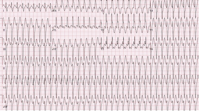
A previously healthy 12 year old presented to the ED with a "fast heart rate" that had started about 1.5 hours prior to presentation. She was reportedly a healthy child and active in several sports. She had a fairly active day and had been swimming off of a boat when her symptoms started. Her mother thought that she was just anxious, so took her home and had her try some deep breathing exercises.
Here is a higher resolution image, but missing V4-V6:
 |
| There is regular monomorphic tachycardia with RR interval of about 0.285 sec (285 ms), for a rate of 210 bpm QRS duration is about 120 ms. What do you think? |
Comment: This is a wide complex tachycardia (but only minimally wide!).
Differential Diagnosis
NOT atrial fibrillation because it is regular.
NOT polymorphic VT (Torsades de Points, caused by Long QT or Catecholaminergic VT) because all the QRS are identical.
1. SVT with aberrancy
2. AV nodal reciprocating tachycardia (AVRT, antidromic SVT, using an accessory pathway, as in WPW).
3 Ventricular tachycardia (VT)
a. "idiopathic" fascicular VT, which has a structurally normal heart
b. "Standard" VT, which occurs in a structurally ABnormal hearts such as cardiomyopathy
So let's think it through:
1. SVT with aberrancy:
A. Sinus tachycardia (a form of SVT) with aberrancy: Sinus tach can beat this fast in a child. But there are no P-waves.
B. Paroxysmal SVT (PSVT) with aberrancy.
The morphology is similar to RBBB (large R-wave in V1, large S-wave in V6) plus left anterior fascicular block (LAFB) (rS in II, III, aVF, and qR in aVL, which gives it a left axis deviation, away from inferior leads).
Is it PSVT with RBBB + LAFB? Unlikely.
i. There is absence of rsR' in V1, typical of RBBB.
ii. QRS duration of 120 ms is atypical for RBBB.
iii. RBBB Aberrancy implies a refractory right bundle. Although the right bundle is the bundle most likely to be refractory, a child of 12 years should be able to fully conduct at a rate of 210.
Thus, SVT with aberrancy is unlikely.
2. Antidromic AVRT
Antidromic AVRT travels down through the accessory pathway and up through the AV node. After traversing the accessory pathway, it must transmit through myocardium (not through specialized conducting Purkinje fibers), which is slow. Thus, there should be an initial wide depolarization, akin to a delta wave.
If you thought it was AVRT, it would be easy to test with adenosine. AVRT depends on the AV node for its circuit and will be terminated by an adequate dose of adenosine.
3. Standard VT (structurally abnormal heart)
Unlikely in a healthy child who has been playing sports all day. But it is possible to have myocarditis or other unknown cardiomyopathy. Standard VT is rarely this narrow, at only 120 ms, though the age of the patient could narrow it. A bedside ultrasound assessment of would be helpful: normal heart and good contractility would be strong evidence against this diagnosis.
4. "Idiopathic" Fascicular VT (structurally normal heart). ["Idiopathic" VT is not really idiopathic any more, but rather is known to be due to reentry. Most are posterior fascicular VT or right ventricular outflow tract VT (RVOT).]
Posterior fascicular VT is a wide complex tachycardia with RBBB and LAFB morphology. It initiates in the posterior fascicle. A re-entrant rhythm that starts in the posterior fascicle will initiate a rapid depolarization down that fascicle (resulting in a short duration r-wave in inferior leads), then an upward directed depolarization through myocardium (wide) to the anterior-superior LV (resulting in S-waves in inferior leads and an R-wave in aVL). It will also transmit to the right ventricle and result in an RBBB-like morphology.
One must also consider RVOT, right ventricular outflow tract tachycardia. However, the morphology is wrong for RVOT (see below).
A normal bedside ultrasound would be helpful.
Case Progression, as described by the treating physician:
"My initial thought was this was either SVT with aberrancy or V-tach. She was stable with an SBP of 146. Her only complaint was shortness of breath but she was not in any visible distress though she appeared anxious. I asked about family history and she did not have any family history of cardiac problems or arrhythmias. She denied any stimulant use or ingestions."
"The rate was regular so I decided to try adenosine. I did this first with 6 mg then with 12 mg. Nothing happened, not even a decrease in HR, and I thought that this could be V-tach so I called the pediatric cardiologist at Children's who told me I was pushing the medication wrong and that I needed to do it with a three way stop cock. He said to keep upping the dose until the rhythm broke."
"I didn't think this sounded right but I did it anyway (in retrospect I should have insisted he look at the EKG first). When this didn't work he finally looked at the strip and agreed that this could be fascicular V-tach. We discussed transfer and he suggested Verapamil. I did give her a 5mg dose and she rapidly converted (2nd EKG). She remained stable and was transferred to Children's. She will have an ablation next month."
"Can you comment and let me know if I should have been immediately onto V-tach? I don't think Adenosine was the wrong thing to try first. If a patient like this were unstable I would have tried electrical cardioversion. "
"Any thoughts other than this?"
"How did you know when I showed it to you that it was from the posterior fascicle?"
"Any thoughts other than this?"
"How did you know when I showed it to you that it was from the posterior fascicle?"
 |
| Sinus Tach, otherwise normal |
Comment:
I knew when I looked at the ECG that it was fascicular VT because of all the features discussed above. Adenosine certainly will not hurt this, but it will not help either. Had it been AVRT, adenosine would probably have converted it.
Verapamil in VT: One might be squeamish about giving such a strong negative inotrope as verapamil to someone with VT. This is due to the fact that most VT occurs in structurally abnormal hearts, especially in ischemic cardiomyopathy, with a low ejection fraction. Giving verapamil to a patient with VT due to cardiomyopathy could indeed be disastrous.
Thus, our minds associate disaster with verapamil and VT. In fascicular VT, we should not carry this association. Of course, if you are hesitant about giving verapamil, just confirm good LV function (good contractility) with bedside ultrasound.
Just for contrast, here is another "idiopathic" VT. This is a case of right ventricular outflow tract VT (RVOT VT). RVOT is adenosine responsive. I presented the case here.
Both RVOT and posterior fascicular VT occur in structurally normal hearts and are usually very well tolerated.
Fascicular VT from the posterior fascicle is responsive to verapamil. RVOT, by contrast, is indeed responsive to adenosine. Therefore, I would not be surprised it if often gets misdiagnosed as SVT with aberrancy after it converts with adenosine.
Learning Points:
1. Wide complex tachycardia in a child with an otherwise normal heart is likely to be one of the "idiopathic" VTs such as fascicular VT.
2. Fascicular VT (not RVOT) originates in the posterior fascicle, and therefore has RBBB/LAFB morphology.
3. Fascicular VT usually has a relatively narrow QRS (up to 140 ms), whereas VT in a structurally normal heart usually has a wider QRS (not always!).
4. Fascicular VT is verapamil responsive.
5. If you can, ascertain good contractility before giving verapamil.
6. Adenosine will often work for RVOT, which has an LBBB morphology with an inferior axis.


The rhythm is a regular WCT ( = Wide-Complex Tachycardia) at ~ 210/minute without sign of atrial activity. Dr. Smith has reviewed the principal differential diagnosis. I don’t think QRS duration is particularly helpful here, given that the patient is a 12-year old child (for whom normal QRS duration is a bit less than in adults). So the QRS is definitely wide, though not excessively so — but probably overlaps most entities to be considered. What strikes me — is the obvious suggestion of bifascicular block (RBBB/LAHB), though of atypical morphology. In an otherwise healthy child (who does not have congenital heart disease or other structural heart abnormality) — one would expect a more typical RBBB morphology with a clear triphasic appearance with taller right-rabbit ear in lead V1, instead of the atypical picture we see here in V1,V2,V3. Considering this and integrating it with the important Learning Points Dr. Smith emphasizes (ie, that fascicular VT often occurs in otherwise normal hearts) — places Fascicular VT high in the differential. Against WPW here is the lack of a delta wave in a tracing in which at least 9 leads manifest a clearly defined and narrow initial deflection. While professing no expertise in pediatric arrhythmias — my approach would have been identical to that attempted = initial administration of Adenosine (which shouldn’t be harmful, and which may convert some of the entities being considered) — followed by cautious trial of Verapamil (which often works amazingly well to convert fascicular VT). GREAT case with happy ending!
ReplyDeleteIf Verapamil is not available, would Amiodarone be safe/effective??
ReplyDeletePossibly, but not as reliably. Electricity would work.
Deleteso is the problem with verapamil the systolic function only without being sure about the diagnosis ( sure not suggested ) ?
ReplyDeleteI don' t know if this is a smart question but could we use verapamil if the systolic function is good and the vt is caused by electrolyte problems
Thanks
Nice case
Bashar,
DeleteVerapamil will only work for this particular kind of VT. So only use it for this, not for electrolyte-induced VT.
Steve
Great case. I'm a fairly junior doctor, so I try to keep things simple in difficult cases such as this. I was always taught, if it's wide, treat it wide. Even though there is no sign of delta wave, or irregularity (afib with wpw), my general inclination would be to avoid av nodal blocking agents or ccb's. Perhaps just go straight to procainamide? That said faced with this case in front of me, I could see myself convincing myself it's probably just svt with abberancy and giving adenosine x 2. I think my personal preference would have been amiodarone or procainamide, any reason NOT to use those?...verapamil would have made me nervous, as I have not typically used it, and know all too well it's negative inotropic effects (saw 2-3 serious ped's verapamil OD's with small doses).
ReplyDeletethey are simply unlikely to work. If you don't know what a regular wide complex tachycardia is, you can shock it. You don't need to avoid AV nodal blockers unless there is BOTH WPW and atrial fib. Adenosine won't work for this, though it will work for a regular tachycardia due to WPW. Amio and Procaine won't work. Verapamil only dangerous if there is poor LV fct.
Deleteextraordinary case. thank you, Steve
ReplyDeleteThanks, Tom!
Delete