Sunday, April 13, 2025

Chest pain and this ECG. Angiogram totally normal. Is this myocarditis?

A man in his early 30s was walking when he developed central chest pain which was non-radiating, then had a syncopal event with bowel incontinence, and when he woke up he had ongoing chest pain. Notes never having symptoms like this before, pain is so severe its causing SOB.  

He called 911.  Medics recorded a BP of 79/52 with pulse of 47.

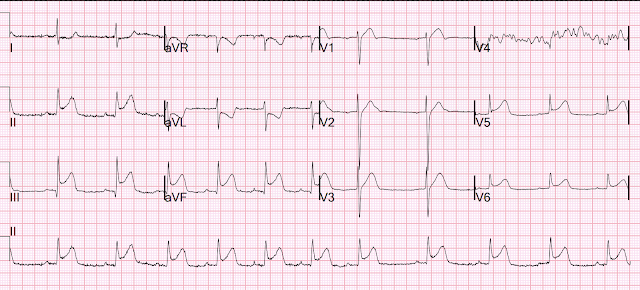
They recorded this ECG: 

Obvious inferior STEMI/OMI
What else?


There is also STE in V1 which is diagnostic of right ventricular OMI in this situation, and partly explains the syncope and hypotension (along with the bradycardia).

A second ECG was recorded by medics:

Now V1 is even more remarkable


So this is an obvious proximal RCA OMI, with inferior and right ventricular OMI.

The cath lab was activated by the medics.

He arrived in the ED and had this ECG recorded:


The first high sensitivity troponin I returned undetectable (<3 ng/L).

The angiogram was normal

The post angiogram ECG is here:

All evidence of OMI is gone.

Formal echo was normal.

I was told of this case the next day and looked for troponins.

The only troponin measured was the first.  

I requested that another be measured, and it returned at 49,000 ng/L

The team thought then that this was myocarditis.

Why can this not possibly be myocarditis?

Because inflammation does not disappear within 2 hours and thus the ECG would not normalize as this one did.

An MRI was done and showed inferior transmural MI.


Here it is annotated in red:


Our extremely smart radiologist, Gopal Punjabi, assures me that this finding can only be due to myocardial infarction, not myocarditis.

The patient was found to have an embolic source.  It was more interesting than that but in the interest of privacy I cannot give more details.


So this is "MINOCA".  Myocardial Infarction with Non-Obstructive Coronary Arteries.

Etiologies (list not comprehensive):

Coronary Spasm.  Provocative testing is very helpful for this

Coronary Thrombus with lysis (one must do optical coherence tomography or at least intravascular ultrasound to find thes non-obstructive plaques that ruptured.

Embolism with lysis.  Need to find an embolic source.  Emboli frequently do not lyse and need to be intervened upon.

Coronary microvascular dysfunction

Coronary dissection


Read more about MINOCA and its etiologies here:

https://www.frontiersin.org/journals/cardiovascular-medicine/articles/10.3389/fcvm.2022.1032436/full


===================================

MY Comment, by KEN GRAUER, MD (4/13/2025):

===================================
Today's case brings up several important points regarding the entity known as, "MINOCA" ( = MI with Non-Obstructive Coronary Arteries)

  • As I wrote in My Comment in the November 16, 2023 post in Dr. Smith's ECG Blog — "I suspect that the entity known as MINOCA is not fully appreciated by many clinicians." I suspect this statement is still true 2 years later, in 2025.

As I wrote in that Nov. 16, 2023 Comment:

  • An estimated 5-15% of patients (depending on the studied population) who are diagnosed with either STEMI or NSTEMI — turn out to have MINOCA, in which cardiac catheterization fails to reveal any infarct-related artery (ie, no "culprit" vessel with at least 50% stenosis) — and no clear systemic etiology is found to explain the patient's presentation to the hospital (Tamis-Holland et al: AHA Scientific Statement on MINOCA — Circulation 139:e891-e908, 2019 — Broncano et al — RadioGraphics 41:8-31, 2021 — and — Sykes et al — Interventional Card Review 16:e10, 2021).
  • As a result of how surprisingly common MINOCA is — We need to know about this entity if we hope to optimally treat the surprisingly large population of patients who have MINOCA.

How to Diagnose MINOCA?
The answer to this question is simple:
  • Is there evidence that an acute OMI has occurred?
  • If so — Was timely cardiac cath "negative" for confirming acute infarction? If so — then the patient has MINOCA.

Regarding Today's CASE:
  • The history in today's case strongly suggests an acute cardiac event: The patient had sudden onset of severe and persistent Chest Pain.
  • The initial Troponin was negative. BUT — as we often emphasize, the initial Troponin may be negative in a significant percentage of acute MIs! The repeat Troponin requested by Dr. Smith removed all doubt about whether there was acute infarction (ie, as repeat Troponin = 49,000 ng/L).
  • Despite the poor resolution of the 2 EMS ECGs — the diagnosis of acute RCA OMI is immediately established by these EMS ECGs until proven otherwise.
  • The cardiac cath was "normal".
  • The Unmistakeable Conclusion: By the definition I suggested above — today's patient manifests MINOCA.

The EMS ECGs:
As per Dr. Smith — the diagnosis of acute inferior STEMI is obvious from the history of new, severe CP in a patient with the initial EMS ECG that I show below (TOP tracing in Figure-1).
  • Rather than trying to "pretty up" today's EMS ECGs with PMcardio — I decided to preserve the originals, albeit with some lightening of the background "noise".
  • I am uncertain about the rhythm in both EMS tracings, other than to say that the rhythm is supraventricular (narrow QRS) — and both slow and at least fairly regular (rate <50/minute)I simply cannot reliably make out atrial activity. That said — awareness that the rhythm in these 2 tracings is fairly regular and supraventricular suffices for identifying the KEY finding (as in the next bullet below).
  • Marked ST elevation in each of the inferior leads — in association with reciprocal ST depression in leads aVL and I — defines this tracing as an inferior STEMI.
  • Acute RCA occlusion is strongly suggested by the presence of ST elevation in lead III>II, and the marked reciprocal ST depression in aVL. In this context (as per Dr. Smith) — the abnormal ST segment straightening, elevation and terminal T wave inversion in lead V1 in the setting of acute RCA OMI (within the RED rectangle) — defines acute RV involvement (and tells us that it is the proximal RCA that is occluded at the time ECG #1 was recorded).
  • Advanced Point: I suspect the ST coving without either elevation or depression in lead V2 — is the result of attenuation of forces (ie, some cancelling out) of the ST elevation that we would have seen in lead V2 from the acute RV MI — with the ST depression that we otherwise would have seen if there was no RV MI, because of associated posterior OMI.
  • P.S.: Given clear indication of acute RV MI on EMS ECG #1 — sublingual NTG should be avoided! And, since a hypotensive BP = 79/52 was recorded by the paramedics — Consideration should be given to the potential need for IV fluids(For more on the ECG diagnosis and consequences of acute RV MI — Check out My Comment in July 19, 2020 post and the August 2, 2024 post).

There is minimal change in the 2nd EMS ECG:
  • The principal difference that I see in EMS ECG #2 — is in the chest leads, where: i) Lead V1 has obviously become more "bulky" (from the acute RV MI)ii) There now appears to be flattening of the ST segment in lead V2, now with terminal T wave positivity (presumably reflecting more activity from the ongoing associated posterior OMI)andiii) Leads V3-thru-V6 all look slighty (subtly) more hyperacute, suggesting associated lateral MI — perhaps reflecting compromised perfusion of postero-lateral branches from the PDA. 
  • None of these subtle changes in the chest leads of EMS ECG #2 alter in any way the conclusion we immediately arrived at on seeing EMS ECG #1 (namely, that there is acute RCA occlusion with acute RV involvement in this acute inferior STEMI ).

Figure-1: Comparison between the 2 EMS ECGs in today's case.

============================================
By way of review — I've copied what follows below from My Comment in the November 15, 2023 post.
  • The importance of the points cited below is highlighted by today's case, in which: i) Consideration of the differential diagnosis for MINOCA (shown in Figure-2) — facilitated identification of an embolic source as the cause of the acute MI in today's case; ii) As per Dr. Smith — the simple clinical reality that the ECG changes of an acute STEMI do not disappear within 2 hours immediately ruled out acute myocarditis as a cause; andiii) Despite acute OMI with spontaneous reperfusion remaining the most common "cause" of MINOCA — sequential evaluation ruled this out as the cause in today's case.


Some Clinical Points about MINOCA:
Because all emergency providers will periodically encounter MINOCA (ie, the 5-15% estimated incidence of MINOCA that I cited above in patients initially diagnosed as having a STEMI or NSTEMI) — I've added Figure-2, which summarizes the more common entities associated with MINOCA.
  • An all-too-common misconception is that the absence of obstructive coronary disease on cardiac catheterization rules out acute coronary occlusion as the cause of the patient's acute event. This is not the case.
  • To paraphrase Dr. Smith's comments in the May 19, 2020 post: — Non-obstructive coronary disease does not necessarily imply no plaque rupture with thrombus. This is because non-obstructive plaques can fissure, thrombose, totally (or near totally) occlude, have autolysis (spontaneous lysis of thrombus with reperfusion) — yet have less than 50% obstruction at angiography. These plaques will often not be recognized as "culprits", because no fissuring or ulcertaion is seen. As a result — determination of plaque disruption in such patients can only be diagnosed by use of intracoronary imaging — with either higher-resolution OCT (Optical Coherence Tomography) or IVUS (Intra-Vascular Ultra-Sound).

  • Bottom Line: Despite lack of obstructive coronary disease on cardiac catheterization — the most common cause of MINOCA is probably still an acute OMI that spontaneously reperfused, and was no longer evident by the time cath was performed.

Additional Points about MINOCA:
I found it surprising to learn that the initial description of acute MI despite normal coronary vessels is not a new concept — having been first described ~80 years ago (with eventual adoption of the term, "MINOCA" in 2013).
  • The 3 most common causes of ACS (Acute Coronary Syndromewithout coronary disease are: i) Myocarditis (up to 1/3 of these patients); ii) Takotsubo cardiomyopathy; andiii) MINOCA.
  • There is a trend toward these patients being younger — with a greater relative percentage of women — and fewer traditional cardiac risk factors.
  • Longterm prognosis of patients with MINOCA clearly depends on the underlying etiology — but it's important to appreciate that this entity is not benign, with similar mortality as for patients with obstructive coronary disease following their infarction.

  • Cardiac MRI — provides an answer to the etiology of patients with MINOCA in more than 2/3 of cases. 
  • Cardiac MRI successfully identifies ~80% of patients with acute myocarditis by picking up evidence of inflammation — with the distinct advantage of being noninvasive compared to endomyocardial biopsy.
  • Use of LGE (Late Gadolinium Enhancement) — is routinely recommended with cardiac MRI to increase diagnostic yield, as a means to identify fibrosis and other abnormalities in cardiac tissues.
  • Cardiac MRI (especially with the addition of LGE) provides insight to longterm prognosis of patients with MINOCA.


Figure-2: Classification of Underlying Diagnoses in Patients with MINOCA (Adapted from Table-1 in Sykes et al: Interventional Cardiology Review: 16:e10, 2021)NOTE: As per Sykes et al — The entities listed under "Other Etiology" may be diagnosed following further investigation and should be considered separately (because they are typically associated with myocardial injury but not considered an MI by the 4th universal definition of MI). This is an important indication for cardiac MRI in patients suspected of MINOCA. 


Friday, April 11, 2025

Look what happens when you advocate for your patient with a skeptical cardiologist.

While at ACC, I received this text message from a former resident:

Good morning Steve! Hope you’re doing well.

Just curious what you think of this?


He writes: "To me, the downsloping ST in V2/V3 is always OMI until proven otherwise. Also looks hyperacute inferior." 

"I activated cath lab, cardiologist is not impressed. Trop pending." 


Smith: "It is diagnostic of OMI!  Cardiologists are rarely impressed because they do not understand OMI ECG findings.  Always advocate for the patient even if the cardiologist disagrees."

The inferior T waves are inflated. They are hyperacute. There is a reciprocally inverted hyperacute T wave and lead AVL.  The slight ST depression in right precordial leads is diagnostic of posterior OMI. Inferior and posterior OMI go together.  It all fits together as an inferior and posterior OMI.


He then provided some history: "Classic story for angina, few hours of crushing chest pain."

The cardiologist said to me: “those aren’t hyperacute T waves and this isn’t a posterior MI, but I’ll come in anyway.”

"He’s here now, and think he’s about to take the patient to the lab, even though he doesn’t believe me." 

"He tried to teach me about the EKG and I just said that you were my mentor and taught me to treat it as an OMI, he still didn’t get impressed but took him to cath anyway." 

"I was using this post (below) to educate my scribe, maybe I’ll send to the cardiologist, if I’m feeling brave" 

7 steps to missing posterior Occlusion MI, and how to avoid them.  

Here is the Queen of Hearts interpretation:


Here is the cardiologist's impression: "EKG does not show a STEMI."


Here is the cath report (TIMI-2 flow in the circumflex -- stented):


Learning Point:

Just because the cardiologist says it is "not a STEMI" and "wants to teach you about the ECG", it does not mean you should back down.

They do not recognize these patterns.

Use the Queen of Hearts!!


Here is another nice example of a posterior OMI:

Occlusion/reperfusion through 6 ‘normal’ ECGs


===================================

MY Comment, by KEN GRAUER, MD (4/11/2025):

===================================
Today's case raises the question of, "What to do when you know you are right — and, you know that your consultant (in this case, the On-Call cardiolgoist) is wrong?"
  • The Answer: The patient comes first — which means that when you know you are right (ie, in this case, when you know the patient is having an acute OMI) — that you should continue to tactfully (but firmly) convey what you know is correct until appropriate management is undertaken (which in today's case was timely cath with PCI ).
  • The "good news" — is that results of a timely cath will almost always reveal the answer (which in today's case confirmed what Dr. Smith's former resident predicted, namely acute posterior OMI).

NOTE:
 Significant delay in performing cardiac cath may sometimes result in a false negative result (ie, If some time has passed and there has been spontaneous reperfusion — and/or if intra-vascular imaging is not done in cases in which standard cath is equivocal) — in which case, the "proof' may reside with Troponin rise and serial ECGs that show evolution of acute changes, ultimately with reperfusion T waves in the affected area(s).

  • For an example of how it can happen that potential false negative cardiac caths occur — Check out the March 17, 2025 post.

What Else To Do When You KNOW You Are Right?
I submit that the "other things" you can do when you know you are right (but your consultant remains unconvinced) — include the following:
  • Show your consultant how QOH (Queen OHearts) may be of assistance! As per Dr. Smith — there was no hesitation by QOH calling today's initial ECG a STEMI Equivalent that merits an "immediate invasive strategy".
  • Tactfully (but firmly) convey to your doubting consultant that in a case such as today's, in which this patient presents with a "classic story for angina, with a few hours of crushing CP" (Chest Pain) — You see ECG abormalities in no less than 10/12 leads, that when put together, tell a storyHopefully — it will be hard for your doubting consultant to negate your story when cardiac cath proves your prediction to be completely correct.

What are the ECG Findings?
For clarity in Figure-1 — I've reproduced and labeled today's initial ECG. The rhythm is sinus bradycardia at ~55-60/minute.
  • My "eye" was immediately captured in this patient with new crushing CP — by the ST-T wave appearance in lead V3, supported by the abnormal ST-T wave in neighboring lead V2 (the leads within the RED rectangle). As per Dr. Smith's former resident — in this patient with new crushing CP, the diagnosis of acute posterior OMI can be made within less than 5 seconds — unless and until proven otherwise by cardiac cath.
  • The Mirror Test in lead V3 is clearly positive (See My Comment at the bottom of the page in the September 21, 2022 post). But regardless of whether or not you are a "fan" of the Mirror Test — Dr. Smith's "mantra" is all that is needed = maximal ST depression in one or more of the leads V2,V3,V4 in a patient with new CP = posterior OMI until proven otherwise.
  • Abnormal ST-T wave appearance continues in neighboring chest leads V5,V6 (both of which show ST straightening with slight depression and disproportionate terminal T wave positivity).
  • As always — whenever we see evidence of acute posterior OMI — we should look closely for associated acute inferior OMI (ie, While we'll occasionally see isolated posterior OMI — the common blood supply to inferior and posterior LV walls most often results in simultaneous acute inferior and posterior lead changes).
  • Each of the inferior leads in today's initial ECG manifest ST segment straightening, with disproportionately taller and "bulkier"-than-expected upright T waves that we know are hyperacute because: i) These T waves are very large considering small QRS amplitude in these same leads; ii) These inferior lead T waves are seen in association with acute posterior OMI in the chest leads; andiii) There is reciprocal ST depression in lead aVL (and to a lesser extent — in the form of abnormal ST segment flattening in lead I, which is the other high-lateral lead).

BOTTOM Line: Credit to Dr. Smith's former resident for immediately recognizing today's acute infero-postero OMI, with need for prompt cath. Credit to this clinician for importantly advocating for his patient — who as a result, received timely PCI that was needed. 


Figure-1: I've labeled the initial ECG in today's case. (To improve visualization — I've digitized the original ECG using PMcardio).


Wednesday, April 9, 2025

Anterior OMI with RBBB has VF x 3: how to prevent further episodes of VF?

A middle-age woman with no previous cardiac history called 911 for chest pain.

This was her prehospital ECG:

What do you think?


There is sinus rhythm with RBBB and obvious LAD OMI (proximal LAD occlusion): hyperacute T-waves in I, aVL and minimal STE in V1, V2.
This is diagnostic of Acute LAD OMI

In case you are wondering what the Queen thought, here she is:
Notice that she also diagnoses Low Ejection Fraction.

The paramedics diagnosis was "Possible Anterolateral STEMI."  I don't know what the device algorithm interpretation stated.

I am not certain if there was a prehospital cath lab activation, but there should be.

On arrival in the ED, a bedside ultrasound showed poor LV function (as predicted by the Queen of Hearts) with diffuse B-lines.   

Initial BP was 120/96, HR 102, SpO2 98%. and this was recorded:

RBBB with proximal LAD Occlusion is very high risk.  

When this came off the machine, I turned to the resident in charge and whispered "This is very bad."


3 seconds later, she went into ventricular fibrillation.

We rapidly defibrillated her, and with return of normal sinus rhythm.

Several minutes later the patient developed V-fib again > 200J defibrillation with return to NSR.

Rapid sequence intubation was performed for airway protection in setting of recurrent V-fib and defibrillations. 

Chest X-ray also showed pulmonary edema.

The patient then had 2 subsequent episodes of V-fib requiring defibrillation, with return to NSR.

She was given 2 mg Magnesium.  Potassium was 4.5 mEq/L

While waiting for the cath team, and to prevent further episodes of VF,  lidocaine 100 mg administered followed by 50 mg q5 min x 3 afterwards (for a total of 250 mg over 15 minutes), followed by a lidocaine infusion of 3 mg/min

Lidocaine dosing: Lidocaine rapidly redistributes from the extracellular to the intracellular space.  A high extracellular (serum) level is toxic.  One must give it in serial boluses to give it time to redistribute.  Thus we gave 1.5 mg/kg, followed by 0.75, then 0.75, then 0,75 spaced 5 minutes apart.  Furthermore, it is rapidly metabolized and therefore an infusion is necessary to maintain adequate therapeutic levels.

The patient was transported to cath lab accompanied by the resident, with BP 50/30 en route improved with a push dose of 100 mcg epi.

She had no further episodes of VF.

Angiogram:

2. LAD: type III-IV vessel with a proximal thrombotic or embolic occlusion

(TIMI 0 flow).  The final angiographic result is very good.


Troponins

Initial troponin was 24 ng/L (barely above URL).  More proof that a huge STEMI may have normal or near normal initial troponin.   We showed this in this article in JAMA Cardiology.

Peak troponin was greater than 60,000 ng/L -- too high to measure.


Echo 9 months later:

Enlarged left ventricular cavity size (LVEDD 6.2 cms) with moderately reduced systolic function. Left ventricular cavity visually appears spherically remodeled.  The estimated left ventricular ejection fraction is 32%.  Akinesis of mid to distal anterior wall and anterior apical, lateral apical, inferior apical, and septal apical walls.


ECG many months later:

RBBB with well-formed Q-waves in V1-V6, also I, aVL.  Very large old anterolateral MI.

In spite of rapid reperfusion, there was a lot of myocardial damage.


Now, many months later, the Queen correctly diagnoses reperfused OMI and low EF (as well as RBBB and LAFB of course)


Why did I use lidocaine?

See reference below.  Lidocaine is effective at preventing even the first episode of VF during acute MI.  I do not give it in all, however.  I give it when there has already been one or more episodes of VF.  Why not amiodarone?  1) as far as I can tell, there is very little data on amiodarone for this indication  2) amiodarone has beta blockade effects which could be deleterious in a patient with large anterior MI with pulmonary edema and at risk for cardiogenic shock (and she did go into shock. 

Lidocaine does not have negative inotropic or chronotropic effects.

_________________

Lidocaine as prevention of further episodes of Ventricular Fibrillation.

REFERENCE

Prevention of primary ventricular fibrillation in acute myocardial infarction with prophylactic lidocaine


Primary ventricular fibrillation (VF) during an acute myocardial infarction (AMI) occurs with a high incidence and mortality rate with or without thrombolysis. The incidence varies from 2% to 19% depending on the definition of “primary.” Primary VF in this study refers to fibrillation occurring in the absence of shock or pulmonary edema. Mortality rate, when primary VF occurs, is 2 to 4 times greater than when it does not. 

Lidocaine had been used for the prevention of VF since the 1960s after coronary care units became a standard setting for the treatment of AMI.7 

Smith comment: Yes, believe it or not, when I started out in the mid 1980's, every patient with acute MI was started on a lidocaine drip!  I'll never forget when I ordered such an infusion in 1991 and then my patient started seizing and I looked up and the nurse had hung the lidocaine wide open!

However, the 1996 guidelines of the American Heart Association and the American College of Cardiology (AHA/ACC) for the treatment of AMI considered prophylactic lidocaine a class III indication.8 This recommendation was based on 4 meta-analyses that hypothesized that the increased mortality rate among patients who received lidocaine was due to asystole or atrial ventricular or sinoatrial block, although without supporting data.

Prevention of VF has been impeded by the publication of the 1996 recommendations of the American Heart Association and American College of Cardiology against the use of prophylactic lidocaine based on meta-analysis studies implying toxicity. This observational study of 4,254 patients with AMI reports the incidence and mortality rates of primary VF over 32 years. Of the 4,254 patients, 4,150 received prophylactic lidocaine, and 104 patients did not receive prophylactic lidocaine due to the 1996 guidelines, after which administration of prophylactic lidocaine was governed by physician choice. The incidence of primary VF was 0.5% among the 4,150 who received prophylactic lidocaine and 10% among the 104 who did not due to physician choice (p <0.0001). 

After 1996, 104 patients did not receive prophylactic lidocaine at the discretion of their physician after the publication of the 1996 AHA/ACC guidelines.8 During this 6-year period, 798 patients with AMI were treated, 13% of whom did not receive prophylactic lidocaine.

Among the 4,150 receiving prophylactic lidocaine, sinoatrial block occurred in 0.5% and complete infranodal atrial ventricular block occurred in 0.2%, all secondary to the site of infarction (concurrent serum lidocaine levels were <4g/ml). Asystole was an agonal rhythm in 4%; these patients had been off lidocaine for 48 hours. Mortality rates were 10.5% in patients without primary VF and 25% in patients with VF (p <0.001). Thus, prophylactic lidocaine markedly decreased the incidence of VF in 4,150 patients with AMI to 0.5% compared with trials before and after thrombolysis (2% to 19%) and with the 104 patients in this study who did not receive prophylactic lidocaine (10%). No


Lidocaine was given as a 75-mg bolus over a 90-second interval followed by a 2-mg/min continuous infusion. A second bolus of 50 mg followed in 5 minutes. The infusion was discontinued after 24 hours if ectopic ventricular beats were not observed. If breakthrough ventricular arrhythmias occurred, additional 50-mg boluses were given every 5 minutes, as needed to a maximum of 325 mg. 

Discussion

Five conclusions have been drawn from the results of this and other studies: (1) the incidence of primary VF before and during the thrombolytic era is unchanged and continues to be excessive; (2) primary VF is unpredictable; (3) when primary VF occurs, it portends poor outcomes in morbidity and mortality rates; (4) lidocaine is effective for preventing primary VF; and (5) lidocaine is not associated with an increased incidence of conduction defects when given properly.

__________________________


Another less relevant study

This was a recent study which excluded patients who received anti-dysrhythmics only after ROSC, so not nearly as relevant.  But it does show a strong association of better outcome with lidocaine vs. amiodarone.

Smida T et al.  A retrospective 'target trial emulation' comparing amiodarone and lidocaine for adult out-of-hospital cardiac arrest resuscitation.  DOI: 10.1016/j.resuscitation.2025.110515


Abstract
Objective: The administration of amiodarone or lidocaine is recommended during the resuscitation of out-of-hospital cardiac arrest (OHCA) patients presenting with defibrillation-refractory or recurrent ventricular fibrillation or ventricular tachycardia. Our objective was to use 'target trial emulation' methodology to compare the outcomes of patients who received amiodarone or lidocaine during resuscitation.
Methods: Adult, non-traumatic OHCA patients in the ESO Data Collaborative 2018-2023 datasets who experienced OHCA prior to EMS arrival, presented with a shockable rhythm, and received amiodarone or lidocaine during resuscitation were evaluated for inclusion. We used propensity score matching (PSM) to investigate the association between antiarrhythmic and outcomes. Return of spontaneous circulation (ROSC) was the primary outcome. Secondary outcomes included the number of post-drug defibrillations and survival to hospital discharge.
Results: After application of exclusion criteria, 23,263 patients from 1,707 EMS agencies were eligible for analysis. Prior to PSM, 6,010/20,284 (29.6%) of the patients who received amiodarone and 1,071/2,979 (35.9%) of the patients who received lidocaine achieved prehospital ROSC. Following PSM, lidocaine administration was associated with greater odds of prehospital ROSC (36.0 vs. 30.4%; aOR: 1.29 [1.16, 1.44], n = 2,976 matched pairs). Lidocaine administration was also associated with fewer post-drug defibrillations (median: 2 [0-4] vs. 2 [0-6], mean: 3.3 vs. 3.9, p < 0.01, n = 2,976 pairs), and greater odds of survival to discharge (35.1 vs. 25.7%; OR: 1.54 [1.19, 2.00], n = 538 pairs).
Conclusion: Our 'target trial emulation' suggested that lidocaine was associated with greater odds of prehospital ROSC in comparison to amiodarone when administered during resuscitation from shock refractory or recurrent VF/VT.


===================================

MY Comment, by KEN GRAUER, MD (4/9/2025):

===================================
Dr. Smith insightfully noted on seeing the initial ED ECG in today's case, "This is very bad ...".  I focus my comment on expanding as to why this is so.
  • For clarity in Figure-1 — I've reproduced and labeled this initial ED ECG.

Findings on the Initial ED ECG:
The main difference between the prehospital ECG (shown above in Dr. Smith's discussion) — and this initial ED ECG in Figure-1 — is the presence of 2 PVCs in the long lead rhythm strip.
  • Confession: I remain confused by the literature I have read as to whether (and if so, by how much) risk is increased by the R-on-T phenomenon (ie, in which the R wave of a PVC falls on the so-called "vulnerable period" — that corresponds to the ascending part of the preceding T wave). First observed almost 100 years ago — we now appreciate that while R-on-T PVCs is not a common phenomenon — its occurrence has been shown to precede VT/VFib in a variety of situations, including acute coronary occlusion.
  • For clarity — I've labeled the peak of the T wave with GREEN arrows for a number of sinus-conducted beats in the long lead II rhythm strip of Figure-1. Note that the 1st PVC in this rhythm strip clearly occurs on the peak of the preceding T wave ( = the R-on-T phenomenon), thereby increasing potential risk of precipitating VT/VFib in today's patient (In contrast — the 2nd PVC = beat #11, occurs later in the cycle, and is not an R-on-T).

There is Low Voltage:
As per My Comment in the December 13, 2023 post — the recognition of low voltage (either isolated to the limb leads — or in both limb and chest leads) — should prompt consideration of a differential diagnosis.
  • Despite RBBB — Today's initial ED ECG clearly shows diminished QRS amplitudes in each of the limb leads (ie, <5 mm in all 6 limb leads) — as well as low voltage in all chest leads except for lead V3.
  • Among the causes of Low Voltage that I list in this Dec. 13, 2023 post is myocardial stunning = a transient marked reduction in cardiac contractility, that occurs in response to a major acute insult such as cardiac arrest, after a sustained tachyarrhythmia — or a large acute MI.

Acute LAD Occlusion:
As per Dr. Smith — the patient's initial tracings are diagnostic of acute LAD OMI.
  • In addition to sinus rhythm with the 2 PVCs that we see in ECG #2 — there is RBBB (in the form of an rSR' or QR in lead V1 — with predominant positivity of the QRS continuing in leads V2,V3).
  • Although difficult to determine if there is an initial Q wave or r wave in lead V1 — a definite small q wave is seen in lead V2, with infarction Q waves continuing in the remaining chest leads. Small q waves are also probably present in leads I,II,aVL.
  • Considering the small size of QRS complexes — there is marked ST elevation in leads I,aVL; and in leads V1,V2 — with ST coving in lead V3 — and ST elevation and/or hyperacute T waves in V4,V5,V6. (Remember that the ST-T wave in right-sided leads V1,V2 should be negative with RBBB — and instead the ST-T wave is markedly elevated in these leads).
  • There is marked reciprocal ST depression in each of the inferior leads.

2 More Interesting ECG Findings:

  • T-QRS-D (Terminal QRS Distortion) is seen in lead V2 (for illustration of T-QRS-D — See My Comment in the November 14, 2019 post).
  • Finally — ECG #2 provides a nice example of how PVCs may sometimes show diagnostic ST elevation (seen in lead aVL for beat #5). This is a helpful PEARL to be aware of — since on occasion, acute ST-T wave changes of acute MI may only be seen in PVCs, and not in the rest of the tracing (See My Comment in the October 8, 2018 post).

Impression: Putting the above observations together — the ECG findings that suggest higher risk for this initial ED tracing include:
  • The R-on-T phenomenon (for beat #4).
  • Diffuse low voltage, which given the clinical setting suggests myocardial stunning.
  • An initial ECG with RBBB and ST-T wave abnormalities in virtually all 12 leads, in which there is marked ST elevation and reciprocal ST depression — and in which infarction Q waves are already seen in at least 8 leads. Taken together, these findings suggest an ongoing extensive antero-lateral STEMI

Figure-1: The initial ECG obtained in the ED (which is the 2nd tracing in today's case — obtained after the prehospital ECG that Dr. Smith shows above)


===========================
Reflections on Lidocaine:
Among the first of my academic publications was a manuscript in which I asked the clinical question, "Should Prophylactic Lidocaine be Routinely Used in Patients Suspected of Acute MI?" (J Fla Med Assoc 69:377-379, 1982).
  • This was before thrombolytics gained full use in our admitting hospital. As a result — I got to witness first-hand how the incidence of primary VFib appeared to dramatically decrease in our ICU as a result of the new practice at that time of using prophylactic Lidocaine on acute MI patients.
  • I later got to trace the evolution of Lidocaine use in the series of ACLS books that I wrote — to which I dedicated a full chapter on Lidocaine pharmacokinetics in my 1st edition (1984) — with emphasis on this drug continued in my next several editions, until the progressive changeover in favor of IV Amiodarone — such that by the time of the last edition I wrote (in 2013), Lidocaine was essentially relegated to a 3rd-line agent for VT/VFib.
  • To Emphasize: Even with widespread use of ACLS Guidelines in years past — clinical practice on the emergency use of antiarrhythmic medication is subject to variation, depending on clinician experience and practice, as well as regional use patterns. Add to this the sobering clinical reality of how difficult it is to get objective, controlled, prospective data in the emergency situation of cardiac arrest and life-threatening arrhythmias.

The above said — today's case as described by Dr. Smith illustrates the highly effective use of IV Lidocaine boluses, followed by IV Lidocaine infusion that successfully abolished what otherwise would probably have developed into refractory VFib.


Recommended Resources