Tuesday, June 17, 2025

Anteroseptal OMI? Accurate ECG interpretation is essential to correctly interpreting the angiogram.

Written by Willy Frick

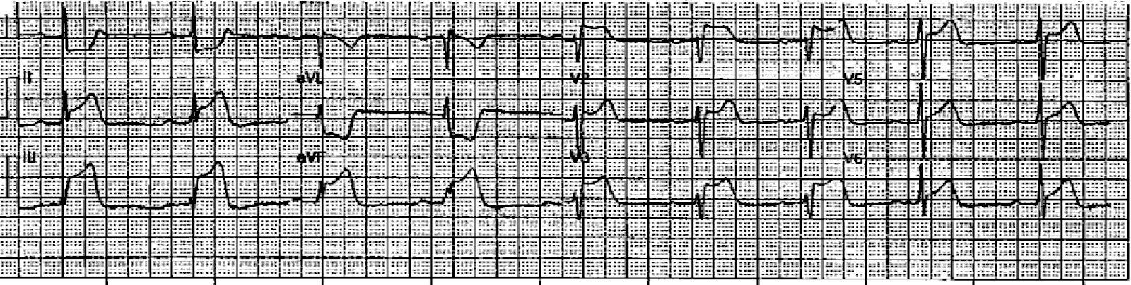
A man in his early 40s with prior anterior OMI s/p bare metal stent to LAD in 2014 presented with acute chest pain, nausea, dyspnea, and diaphoresis. He was brought in by EMS. His first ECG is shown:

ECG 1
Obvious STEMI (+) OMI. What would you guess is the culprit? 


There is STE in V1 and V2 with lateral STD, a pattern known as precordial swirl, so...it should be LAD, right? Sure, there's evidence of ischemia in lead III (and aVF to a lesser degree), but many patients have a long "wraparound" LAD that supplies the distal inferior wall producing inferior STE when occluded.

I sent this ECG to several different friends separately, and everyone said it was suspicious for proximal RCA occlusion with right ventricular infarction. This may be a surprise to many, who are used to thinking of V1-V6 as the anterolateral leads. And used to thinking of RCA as supplying the inferolateral wall.

But remember that V1-2 especially are rightward looking leads. Think of this image:

Figure 1: Schematic representation of V1 and V2 electrodes

This is a transverse CT. The four cardiac chambers are labeled (LA, RA, LV, RV). The right ventricle is the most anterior chamber, and V1 and V2 electrodes are overlying. This is why we see:
NanashÄ« educated me about the term "pseudoanteroseptal" injury pattern of proximal RCA occlusion (since embarrassingly, I had not read this post). For me, the key to this ECG is the STE maximal in V1 indicating a very rightward vector of injury. This is corroborated by STE in III, whereas there is STD in II.

Because of the obvious presentation and STEMI (+) ECG, the patient was immediately taken to cath lab. In fact, everything was highly efficient, and exactly 20 minutes after he arrived in the hospital, he had his first angiogram!


Figure 2: LAO cranial angiogram, annotated on the left, unedited on the right

(Note: This annotation required looking at several different views for me to determine vessel course. And the white arrow in this case is not pointing to an RV marginal, I just ran out of high contrast colors.) The arrow is pointing to the ostium of a bifurcating diagonal branch with a severe lesion felt to be the culprit.

But, does diagonal culprit fit with the ECG? 

Answer: No. 

Our differential was most likely proximal RCA versus possible LAD due to the finding of precordial swirl with massive STE in V1. If LAD produces a swirl pattern, it should be due to involvement of the septal perforators. The diagonal vessel does not give septal perforators, so it should not cause swirl. Diagonal culprits classically cause South African Flag pattern with STE in I, aVL, and V2 with reciprocal STD in III.

The cardiologist then worked meticulously on the diagonal, which was a difficult intervention. The vessel was difficult to wire and required multiple attempts. There were also several other technical challenges including difficulty passing the intravascular ultrasound probe. Additionally, the ostial location of the lesion adjacent to a pre-existing LAD stent (not well seen in the angiogram) added complexity. Although the initial ballooning was quite rapid and well within the 90 minute target, the final post-intervention angiogram of the diagonal vessel was about two and a half hours after the patient arrived.

At that point, the first injection of the RCA was performed.

Figure 3: TIMI 0 proximal RCA

Unfortunately, this image of the culprit was taken 152 minutes after arrival. I made a video explaining the post intervention angiogram for those who are interested.


So although the recorded "door to balloon time" is 43 minutes, this number describes an irrelevant consideration -- time to revascularize a non-culprit lesion. Considering the RCA as the culprit, the door to balloon time was instead 157 minutes.

Echocardiogram showed mild inferior hypokinesis with overall preserved LVEF, 60%. There was evidence of RV dysfunction. (For the echocardiographically inclined, TAPSE was 1.4 cm.)  Troponin prior to the cath was within the reference range, and there was no repeat measurement. Fortunately, the patient did well clinically in spite of the delay.

Learning points:
  • For OMI with STE maximal in V1, RV infarct is likely
  • STEMI metrics frequently conceal missed opportunities for process improvement

Here is a related Case, explained by video:


Right Ventricular MI seen on ECG helps Angiographer to find Culprit Lesion

This is a video I made a while back.  I thought it was worth a re-post.  For those who don't have time to watch a video, you'll have to read the ECG as shown on this still frame because I lost it and cannot post it.

See down below for explanation if you don't want to watch the video.
 
There is inferior STEMI.  But there is also ST elevation in leads V1 and V2.  When you see this, think right ventricular (RV) MI.  The hypotension is further evidence for RV MI.  There was no right sided ECG.

I heard about the case, and saw the ECG, shortly after the patient left for the cath lab.  I called the interventionalist while the patient was on the table and he told me that the occlusion was not in the proximal RCA, but further down.  I asked if he was sure about this, because the ECG would indicate a proximal RCA occlusion with RV MI.  He took another look and realized that the culprit was indeed in the proximal RCA and that the thrombus had embolized distally.  And so he put the stent in the proximal RCA.

Learning point: Even when you have an angiogram, the ECG findings make a difference.

More Cases of Pseudoanteroseptal MI:


===================================

MY Comment, by KEN GRAUER, MD (6/17/2025):

===================================
Several important messages are conveyed by today's case:
  • In a patient with new symptoms — ST elevation in lead V1 merits inclusion of acute RV MI in the differential diagnosis. As per Dr. Frick — the principal differential diagnosis of today's initial ECG (that I've reproduced in Figure-1) is: i) Acute proximal RCA occlusion with RV MI; — vs — ii) Acute proximal LAD occlusion ("Swirl" pattern).
  • "A picture is worth 1,000 words". Acute RV MI becomes especially likely when the ECG looks like the tracing shown in Figure-1 because: i) Chest lead ST elevation is limited to leads V1 and V2, in which the amount of ST elevation is marked in lead V1 (more than 6 mm!) — greater in lead V1 than in V2 — and not seen beyond lead V2 (whereas LAD OMI usually has ST elevation in additional anterior leads); — andii) The ST elevation with Q wave and terminal T wave inversion in lead III (but not in the other inferior leads) indicates associated inferior OMI — and — a rightward ST elevation vector consistent with RV MI.
  • The combination of acute inferior OMI and RV MI implicate the proximal RCA (Right Coronary Artery) as the "culprit" artery.
  • When the possibility of acute RV MI is raised — Get right-sided leads! This apparently was not done in today's case.
  • Awareness of the likelihood of acute RV MI based on the ECG in Figure-1 (and therefore of the proximal RCA as the likely "culprit" artery site) — provides an excellent example of how superior ECG interpretation can aid the angiographer in knowing where to look for the site of acute occlusion. Had the significance of the above-described ECG findings been recognized — myocardial-preserving PCI could have been accomplished 2 hours sooner than it was.
  • Elsewhere on ECG #1 — diffuse ST depression + ST elevation in lead aVR (albeit to a lesser degree than that seen in leads V1,V2) — is consistent with DSI (Diffuse Subendocardial Ischemia) and this patient's history of underlying coronary disease (ie, his prior LAD stent in 2014).
  • Finally — the failure to recognize the probable "culprit" artery from the ECG in Figure-1 led to initial wrong-vessel intervention in today's case — which as per Dr. Frick, led to misclassification of "door-to-balloon" time as "appropriate" in chart records (whereas "door-to-balloon time" of the correct culprit artery was significantly delayed).

Figure-1: The initial ECG in today's case.


Sunday, June 15, 2025

A woman in her 50s with acute chest pain

 Written by Pendell Meyers


A woman in her 50s with no significant past medical history experienced acute anterior chest pain that woke her from sleep and radiated to her back. She described it as "stabbing", 8/10, constant, and associated with nausea. She denied preceding symptoms or recent illnesses. 

Here is her triage ECG during active pain:

What do you think?


This ECG was recorded 30 minutes later with ongoing pain:


Queen of Hearts says no signs of STEMI or Equivalent for both:


New PMcardio for Individuals App 3.0 now includes the latest Queen of Hearts model and AI explainability (blue heatmaps)! Download now for iOS or Android.  (Drs. Smith and Meyers are shareholders in Powerful Medical.)


The physician was worried about possible acute coronary occlusion, and activated the cath lab.

The angiogram showed no significant coronary disease:


First troponin I was 10 ng/L.

Second troponin was 23 ng/L. None further were measured during this hospitalization.

Echocardiogram showed normal EF, no wall motion abnormalities, no pericardial effusion.

_____

There is a lot of data now to show that the Queen reduces false positives by a lot.


2. Midwest STEMI Consortium: decreased false positives by 33% (presented at ACC Quality Summit San Antonio 2024)

3. Willy Frick, from St. Louis University, presented at Heart Rhythm Society: 1 month of chest pain in the ED.  528 chest pain patients, 14 with culprits lesions.  Queen detected 11/14 with zero false positives.  Cardiologist detected 6/14 with 3 false positives.

4. Macherey-Meyer S.  Presented at German Society of Cardiology.  1770 chest pain patients.  56 OMI.  Specificity 0.967.  Negative predictive value 98.4% and Positive predictive value was 34.1%.  Subgroup analysis of patients ruled-out following 0/1h hs-cTnT algorithm identified misclassification as false-positive in 1% of ECGs by AI model. Specificity and accuracy both were 0.989 (95% CI: 0.983-0.994, see table 2). Negative predictive value was 100% (95% CI: 100-100).

There is more unpublished work which shows dramatic decreases in false positive using the PMCardio Queen of Hearts AI Model.

_______


Here is the ECG the next day:

(QoH gets this one wrong, calls it STEMI/OMI, understandably based on the STE in III with STD in aVL.)


She was given colchicine and discharged.


She returned with similar symptoms about 1 month later. 


Repeat echocardiogram at this visit revealed a small pericardial effusion.

 
Cardiac MRI was read as:
Acute pericarditis with signs of early constrictive physiology. The pericardium is diffusely thickened. There is diffuse late gadolinium enhancement of the parietal and visceral pericardial layers consistent with pericardial inflammation. There is a small circumferential pericardial effusion. There are no signs of tamponade physiology. There is ventricular interdependence as demonstrated by mild septal bounce, and the IVC is dilated to 2.9 cm with less than 50% collapse with inspiration - these findings suggest early constrictive physiology. There is no myocardial LGE to suggest inflammation, infiltration, or infarction of the LV myocardium.  


Two troponins were drawn, both less than 6 ng/L.

She was given an extended course of colchicine and ibuprofen. 

Rheumatologic and infectious workups were negative. 


This ECGs was recorded on day 4 of the hospitalization:


This ECG was recorded on day 6 of hospitalization:


Her diagnosis was recurrent idiopathic pericarditis. She was discharged. 


Here is her ECG 6 months later, at an outpatient visit with no active symptoms:


The "four stages of pericarditis on ECG" are typically taught as:


Stage 1: wide spread STE and PR depression
Stage 2: ST segment return to baseline with T wave flattening
Stage 3: T wave inversion
Stage 4: Normalization

Of course, these stages may not follow a typical time course, may not have serial ECGs timed correctly to catch each stage, etc. 

However, this patient seems to fulfill the classic sequence as shown in her second admission.


Please understand that actual pericarditis is incredibly rare compared to normal variant baseline ST elevation.

And please be careful: the harm of one delayed diagnosis of OMI, incorrectly diagnosed as pericarditis, far outweighs the benefit of quickly making many true positive diagnoses of pericarditis. OMI is common, while pericarditis is rare. As Dr. Smith says, "you diagnose percarditis at your peril!"


See these cases if you don't understand how dangerous it can be:


See our other cases involving pericarditis here:


Severe Atypical Chest Pain in a Young Woman: Series of Pericarditis ECGs


Here are some ECG cases of myocarditis in young people:

https://hqmeded-ecg.blogspot.com/2019/06/a-20-something-male-with-acute-chest.html —
https://hqmeded-ecg.blogspot.com/2017/11/anterior-st-elevation-with-elevated.html —
https://hqmeded-ecg.blogspot.com/2017/03/a-young-man-with-sudden-chest-pain.html —
https://hqmeded-ecg.blogspot.com/2014/11/a-young-woman-with-chest-pressure-and.html —
https://hqmeded-ecg.blogspot.com/2014/06/a-29-year-old-male-with-pleuritic-chest.html —
https://hqmeded-ecg.blogspot.com/2013/10/inferior-and-lateral-st-elevation.html —

8 yo with myocarditis:
https://hqmeded-ecg.blogspot.com/2014/05/an-8-year-old-with-syncope-abdominal.html —

16 yo with acute MI:
https://hqmeded-ecg.blogspot.com/2014/05/a-16-year-old-girl-has-syncope-while.html —


===================================

MY Comment, by KEN GRAUER, MD (6/15/2025):

===================================
Medicine is humbling. And while I definitely included acute pericarditis in my differential diagnosis when I first saw today's case — I did not think pure pericarditis (without a component of myocarditis) was likely. Instead — I thought this patient had acute LCx occlusion.

  • For clarity in Figure-1 — I've reproduced today's initial ECG.

Why I Thought:  "Acute MI Until Proven Otherwise" ...
The ECG in Figure-1 shows sinus rhythm at ~90/minute — normal intervals and axis — no chamber enlargement — and lots of ST elevation.
  • This 50-something woman was awakened from sleep by new, severe CP (Chest Pain) — which prompted her ED presentation. As per Dr. Meyers — statistically, acute MI is abundantly more common than acute pericarditis in this setting (and potential morbidity and mortality from missing acute MI far exceeds that for missing acute pericarditis). As a result — Assume acute MI until proven otherwise!

Regarding ECG findings in Figure-1 that suggest acute MI until proven otherwise:
  • Q waves are present in multiple leads in ECG #1 — and these are the same leads that show ST elevation.
  • The ST elevation seems to correlate with acute infero-lateral infarction (I suspected acute occlusion of a dominant LCx ... ).
  • Lead aVL seems to show reciprocal ST changes (albeit the amount of ST depression in aVL is not as much as I'd normally expect, given the amount and acuity of the ST elevation in lead III ).
  • More than just ST elevation — lead V3 (and to a lesser extent, lead V4look hyperacute (BLUE arrow highlighting the disproportionately "fat" T wave peak and "bulkier"-than-expected T wave dimensions in this V3 lead).
  • I initially thought there was T-QRS-D (Terminal QRS Distortion) in lead V4. If true — this would all-but-confirm acute infarction (See My Comment in the November 14, 2019 post in Dr. Smith's ECG Blog for more on T-QRS-D). 
  • Bottom Line: While more data was needed — my initial impression of ECG #1 was acute infero-lateral STEMI in progress with need for prompt cath.


Figure-1: The initial ECG in today's case (with magnified view in the 2 inserts below of leads V4 and V6)

======================================
ECG Findings For and Against Acute Pericarditis:
I did not think today's initial ECG was suggestive of pure acute pericarditis. It clearly could be consistent with acute myopericarditis — as the ECG picture of acute myocarditis can manifest virtually anything, and be indistinguishable from the ECG of acute MI. Consider the following:
  • As Drs. Smith and Meyers have repeatedly emphasized in this ECG Blog — You diagnose acute pericarditis "at your peril" — for the simple reason that in an unselected emergency setting, acute MI is so much more common than pericarditis (and the potential morbidity and potential mortality of missing an acute MI is so much greater than for missing acute pericarditis).
  • In favor of the ECG diagnosis of acute pericarditis — is the diffuse ST elevation (seen in leads I,II,III,aVF; and in leads V3,V4,V5,V6).

Against
 the ECG diagnosis of acute pericarditis are the following findings:
  • Diffuse Q waves are seen in almost all leads in which there is ST elevation. Although these Q waves are small and narrow: i) These Q waves are indistinguishable from the early Q waves of acute infarction in which ST segments are acutely rising; andii) Although inferior lead "normal septal q waves" may be seen in leads II,III,aVF — this usually only occurs with an inferior axis (which is not present in ECG #1) — and, if the inferior Q waves that we see in ECG #1 were the result of "normal septal q waves" — then we should not also see the Q wave that is present in lateral lead I.
  • One of the most characteristic ECG features of acute pericarditis in my experience — is that the ST-T wave appearance in lead II resembles the ST-T appearance in lead I (vs the situation with acute inferior MI — in which the ST-T wave appearance in lead II is much more similar to what we see in lead III, than it is to the lead I ST appearance).
  • T-QRS-D is not present in lead V4 of Figure-1 (As shown in the magnified insert — When we look closely, the BLUE arrow highlights a definite notch at the J-point that negates the possibility of T-QRS-D).
  • Although the ST/T wave ratio in lead V6 is "positive" for pericarditis (ie, the ST/T ratio = 0.33, which is over 0.25) — I did not feel this was a convincing result because the amount of ST elevation in lead V6 is only 1 mm, and therefore potentially subject to error pending minimal change in the point of reference chosen for measurement (See Figure-3 in My Comment in the June 8, 2022 post for more on the ST-T wave ratio in lead V6 for pericarditis diagnosis).
  • With the "textbook typical" ECG picture of acute pericarditis — ST elevation is seen in all but the right-sided leads (which are leads III,aVR,V1). There should not be suggestion of reciprocal change (as we see in lead aVL of Figure-1) — and there should not be a flat ST segment in lead V2 (as we see in Figure-1).
  • As stated earlier — the ST-T waves in lead V3 (and to a lesser extent, in lead V4) look hyperacute (BLUE arrow highlighting the disproportionately "fat" T wave peak and "bulkier"-than-expected T wave dimensions in this V3 lead). In contrast — with the "textbook picture" of pure acute pericarditis, otherwise "normal" ST-T waves tend to look as if "lifted" above the ECG baseline instead of producing the hyperacute appearance that we see in lead V3.
  • NOTE #1: Acute myocarditis could easily have produced the identical ECG picture that we see in Figure-1 — in which case it would be impossible to distinguish between acute myocarditis vs acute MI on the sole basis of this single ECG. But the completely normal cath — negative Troponins — normal Echo without wall motion abnormality — and normal MRI (without evidence of inflammation) ruled out acute myocarditis.
  • NOTE #2: No mention is made in today's history as to whether a pericardial friction rub was (or was not) present. This is unfortunate (but all-too-common in the cases I routinely see posted on the internet) — because IF a pericardial friction rub is heard, then you have made the diagnosis of acute pericarditis!
  • NOTE #3: No mention was made regarding any potential positional relationship or pleuritic nature of this patient's CP (the CP was instead described as "constant" and "stabbing", and associated with nausea). While a patient's description of the nature of their CP is by definition subjective, with imperfect correlation to textbook description of pericardial pain — there is a tendency for pericarditis CP to be "pleuritic" (increasing with inspiration due to commonly associated pleural inflammation) — and "positional" (commonly relieved by sitting up and exacerbated by lying supine, which increases "stretch" on the inflammed pericardium).

BOTTOM Lines: 
  • I've always believed it important to be humbled on occasion by cases in which the patient "doesn't read the textbook" before seeking emergency care. Clinical reality is not always predictable. Despite including specific chapters on the ECG findings of pericarditis for each of the ECG books that I've written over the years — I simply did not feel today's initial ECG suggested pericarditis over acute MI (See Figures-2-thru-6 in My Comment at the bottom of the May 16, 2023 post in Dr. Smith's ECG Blog for an example of such chapters).
  • Today's case is important because: i) It illustrates completely appropriate indication for prompt cardiac cath to exclude the possibility of acute infarction; — ii) It expands our insight regarding potential ECG presentations of pure acute pericarditis (as well as the diagnostic path for confirming this diagnosis)andiii) It keeps us humble (while reminding us that in the great majority of cases in which a patient presents to the ED with sudden onset of severe, new CP — the diagnosis will be an acute cardiac event instead of acute pericarditis or myocarditis).   


Thursday, June 12, 2025

60-something with h/o Coronary Bypass has acute chest pain

A 60-something with h/o Coronary Bypass called 911 for acute chest pain.

Here is the first prehospital ECG: 

What do you think?


There is minimal ST Elevation in inferior leads which could easily be normal.  However, aVL has minimal STD supportive of the diagnosis of OMI.  More importantly, the ST depression in V2-4 is diagnostic of posterior OMI.  There is a hyperacute T-wave in V6, which is the lateral part of the posterior wall and a common supportive finding in posterior OMI.

The Zoll algorithm impressively stated: ***STEMI***

A paramedic student had the PMCardio AI Queen of Hearts on his phone and this is what it reported:


New PMcardio for Individuals App 3.0 now includes the latest Queen of Hearts model and AI explainability (blue heatmaps)! Download now for iOS or Android.  (Dr. Smith is a shareholder in Powerful Medical.)


On arrival, an ED ECG was recorded:

Still diagnostic

When a patient has severe chronic coronary disease, findings which appear to be acute can sometimes be chronic, so in this patient with h/o CABG (coronary bypass), it is wise to find a previous ECG if possible.  

Here it is:

This confirms that all findings are new.


The cath lab was activated

The interventionalist disagreed with the assessment, but agreed to do the angiogram.  


Angiogram results are complicated because of the CABG:


Severe three-vessel obstructive coronary artery disease, with chronic total occlusion of the mid LAD, total occlusion of the mid RCA, and severe subtotal occlusion and severe disease in the left circumflex coronary artery mid segment and proximal segment, as well as severe disease in the OM itself. Noteworthy that the LAD and the left circumflex coronary artery share 2 separate ostia with very close proximity to the origin of both vessels and severe

disease in both vessels.


Saphenous vein graft to the RCA is a large in diameter with significant conduit 2 vessel mismatch but there was no severe disease in the vein graft and the anastomosis to the distal RCA appeared to be okay.


Patent LIMA to the LAD the LIMA is anastomosed to the mid LAD after the large LAD D1 and LAD D2.


Saphenous vein graft to the obtuse marginal is small in diameter with severe ostial disease and total occlusion in the proximal body of the graft.  This is most likely the infarct-related artery.


Unfortunately, for technical reasons, it could not be stented.


Should thrombolytics be given?


I would say that, if there are no contraindications, yes.  I have never seen a case in which the artery was unable to be opened for technical reasons and so therefore thrombolytics were given instead.  


Peak Troponin I was 22,000 ng/L (Typical of a moderate STEMI)


Formal Echocardiogram


Normal left ventricular cavity size, mildly increased wall thickness and mildly reduced systolic function; estimated left ventricular ejection fraction 45-50%.

Regional wall motion abnormality: mid anterolateral and inferolateral hypokinesis.  

This is the term that is used for a posterior wall motion abnormality -- this confirms posterior OMI.


The EF just 9 months prior was 64%, so there was significant myocardial loss.


Learning Points: 

1. If you cannot open the artery for technical reasons, consider thrombolytics.

2. In the setting of acute chest pain, ANY ST depression maximal in V1-V4 is acute OMI until proven otherwise.  We have proven this.

Exceptions

One should be circumspect (careful, that is, and not jumping to conclusions because the STD maximal in V1-V4 may NOT be due to OMI), when there is atrial fibrillation or abnormal vital signs:

1. Atrial fibrillation

Great example: 60-something: chest tightness, palpitations, and ST depression V1-V3

Other Examples:


Cardiac arrest, defibrillated, diffuse ST depression and ST Elevation in aVR. Why?


2. Shock/Hypotension:


3. Tachycardia


===================================

MY Comment, by KEN GRAUER, MD (6/12/2025):

===================================
Today's case is of a 60-something man with known coronary disease — who called EMS for new-onset CP (Chest Pain).
  • For clarity in Figure-1 — I've reproduced the first 2 ECGs in this case.
=================================

QUESTION:
  — How quickly can you make today's diagnosis?
  • Beyond-the-Core: Has there been a change in the repeat ECG?

Figure-1: Comparison of today's initial EMS ECG — with the repeat ECG on arrival in the ED.


ANSWERS:
The purpose of my comment in today's case is to highlight how quickly the diagnosis of acute posterior OMI should be made from ECG #1.
  • Knowing this patient's age and his history of known coronary disease — immediately places him in a higher-risk group for an acute coronary event.
  • Given this history — it should take no more than seconds to definitively diagnose acute posterior OMI because: i) There is maximal ST depression in leads V2,V3,V4 (RED arrows); and, ii) The shape of this ST depression in these 3 leads looks acute (straightened, "shelf-like" ST segments with terminal T wave positivity). There is a positive "Mirror Test" (See My Comment in the September 21, 2022 post, among many others for more on the "Mirror" Test).

As per Dr. Smith — There is a hint in ECG #1 of ST elevation in the inferior leads, with abnormal ST segment flattening in lead aVL. That said, by itself — this would not be diagnostic because of how subtle these inferior lead findings are — and because the essentially flat ST segment in lead aVL is nonspecific, and not convincing of a reciprocal change.
  • There is also ST depression in lead I of ECG #1. Most of the time with acute inferior infarction — the amount of reciprocal ST depression is usually greater in lead aVL than in lead I. Retrospectively (ie, knowing the cath findings) — this ST depression in lead I more likely reflects this patient's severe multivessel disease.

What about the Repeat ECG?
As stated — the diagnosis of acute posterior OMI (and the need for prompt cath with PCI) — should be immediately made on learning the history and seeing ECG #1. That said — I'd add 2 important points related to ECG #2:
  • Point #1: There has been improvement in the ECG findings that were seen in the initial EMS tracing. Specifically: i) Although ST depression is still present and maximal in leads V2,V3,V4 — the amount of depression, and its acute appearance is less in ECG #2 (less of a "shelf-like" appearance to ST segments — with decreased height and less peaked terminal T wave positivity in these leads)ii) There is no longer ST depression in lead V5; andiii) ST depression is less in lead I.
  • Point #2: There is no mention of whether this patient's CP was less at the time ECG #2 was recorded. This is relevant — because IF this patient's CP had decreased at the same time that acute ECG findings were improving, this would provide further support of an acute cardiac event, as well as suggesting some degree of spontaneous reperfusion.


Recommended Resources