This was just published in JAMA Internal Medicine:
The de Winter Electrocardiogram Pattern Evolving From Hyperacute T Waves
It reminded me that many believe, due to the assertions in the original de Winter's article, that de Winter's waves are stable.
In fact, the title was "Persistent precordial ‘‘hyperacute’’ T-waves signify proximal left anterior descending artery occlusion." The authors based this idea of persistence mostly on their perception, not on a rigorous evaluation involving frequent serial ECGs of all patients with de Winter's T-waves.
But de Winter's waves are not stable.
I have seen at least a dozen cases with de Winter's T-waves and they have for the most part not been stable but rather evolving into 1) hyperacute T-waves (without ST depression) or into 2) ST Elevation, or they appear after initial ST Elevation or hyperacute T-waves.
It is difficult to say if they ALWAYS are in flux because, whenever I see one, I activate the cath lab immediately and the artery is opened without ever recording serial ECGs.
Here is one case of a patient I saw. He was a 30-something with chest pain.
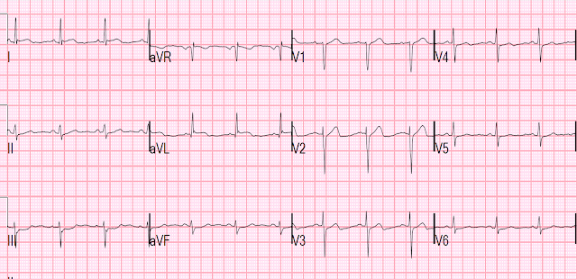
Prehospital ECG:
The cath lab was activated prehospital by the medics.
We recorded another ECG:
30 minutes later, just before arrival of the cath team, we recorded this ECG:
Comment: I frequently am sent ECGs of posterior MI (with anterior ST depression) which are represented as cases of de Winter's T-waves.
Sometimes these are difficult to differentiate, but for the most part, de Winter's waves are far larger (fat and bulky, like hyperacute T-waves) than are the T-wave of posterior MI (which may also have ST depression with an upright T-wave -- we have put 3 such cases way below at the bottom)
Angiogram:
Showed a 100% occluded mid LAD.
Peak troponin I (contemporary) was 101.0 ng/mL (analogous to 101,000 ng/L in a high sensitivity assay and a very large MI).
Echo immediate:
35% EF with anterior, septal, and apical wall motion abnormalities
Echo convalescent, 2 months later:
Better, with EF up to 45-50%
I posted this in 2014:
Is the LAD really completely occluded when there are de Winter's waves?
A male in his 30's complained of sudden severe substernal chest pain. He was rushed to the critical care area where they recorded this ECG:
 Obvious Antero-lateral STEMI due to
proximal LAD occlusion (proximal because it is proximal to first
diagonal, resulting in high lateral STEMI with ST elevation in I, aVL
and reciprocal ST depression in inferior leads)
Obvious Antero-lateral STEMI due to
proximal LAD occlusion (proximal because it is proximal to first
diagonal, resulting in high lateral STEMI with ST elevation in I, aVL
and reciprocal ST depression in inferior leads)The cath lab was activated, and just before going to the cath lab (19 minutes after the first ECG), this ECG was recorded:
I have often wondered if de Winter's T-waves really are due to complete occlusion, or to severe, subtotal occlusion. Perhaps they indicate an open artery with minimal flow and severe subendocardial ischemia, but not total subepicardial ischemia.
Since ACS is so dynamic, with thrombi forming and lysing continuously,
and because the ECG and angiogram are rarely simultaneous, it is
probable that de Winter's T-waves are recorded in a window when the
artery is barely open.
Angiogram:
99% lesion with some flow.
Here is the first post cath ECG, shortly after opening.
Here is the ECG the next day:
Meyers comment: In my shorter experience with de Winter's, I agree that they have always been dynamic. See the following case for a patient with de Winter's T-waves (this one without a lot of ST depression) that held their morphology without evolving "up" or "down" for at least 30 minutes (I recorded perhaps 20 ECGs in attempt to show a reluctant cardiologist progression to STEMI, and they were all identical during this time without any STE or change at all). This is the longest lasting I have ever documented a hyperacute T wave without going "up" or "down." Of course, the ECG after the cath was very much changed. The longest I have seen a hyperacute T wave retain its morphology, without any ST elevation (even subtle STE) is about 30 minutes. It is not at all appropriate to use the word "persistent" for hyperacute T waves in my opinion.
Shoulder pain after lifting a heavy box
de Winter's -- they remained stable for 30 minutes with many ECGs. But we find that this is unusual.
Posterior OMI looks very different from de Winter's
This is a de Winter's that could be mistaken for posterior:
Chest pain and precordial ST depression which resolve, followed by a wide complex rhythm
Posterior
Another posterior OMI -- does not look like de Winter's!!
Meyers comment #2: I was texted the following case's ECG asking "de Winters?" and I said it was instead posterior OMI. I can see why people are confused, but they are very different.
A middle aged man with ST depression and a narrow window of opportunity
This is the ECG recorded after opening the artery of a posterior OMI (after reperfusion). T-waves are somewhat large and upright, but even here should not be mistaken for de Winter's, especially given that it is after opening a proximal circumflex occlusion (supplying the posterior wall). Posterior Reperfusion T-waves.
Also too narrow to be de Winter's


What is the rhythm pre-hospital ECG?
ReplyDeletesinus
Deletesuper cool, super excellent. thank you both.
ReplyDeletetom