This case was written up by Kuan-Yu (Evan) Lin with some edits by Willy Frick. I (Willy) had the pleasure of working with Evan when he was a medical student. Now as an intern, he is exceptional at EKG interpretation because he was able to learn of the OMI paradigm and importance of pattern recognition before getting poisoned by years of learning STEMI.
A 41-year-old South Asian male with history of hypertension, alcohol use disorder and hyperlipidemia, who has a strong family history of CAD presented with central substernal burning, pressure, and pain with associated diaphoresis.
(When seeing a South Asian patient with chest pain, concern for ACS must be heightened, given their disproportionately higher risk of CAD, despite often lacking traditional risk factors.)
Patient initially presented at 9 PM to a referring facility with hsTnI 13 (ref: < 34 ng/L) then 30, then 60. Patient stated the symptoms began that same day at around 3 PM when he was drinking alcohol. He consumed 6 drinks. He denied any history of similar symptoms.
EKGs from the referring facility were not available. Patient was loaded with aspirin, started on heparin gtt, and transferred to PCI capable center for treatment NSTEMI. He was also given GI cocktail and chlordiazepoxide. Patient arrived at 3 AM and the following EKG was recorded, and a repeat high sensitivity troponin T (hsTnT) was 189 ng/L. From chart review, no additional anti-anginals were given but the patient was described as chest pain free.
Lemkes JS, Janssens GN, van der Hoeven NW, et al. Timing of revascularization in patients with transient ST-segment elevation myocardial infarction: a randomized clinical trial. Eur Heart J [Internet] 2019;40(3):283–91. Available from: http://dx.doi.org/10.1093/eurheartj/ehy651
Lemkes et al. randomized 142 patients with transient STEMI (whose symptoms and ST elevation had resolved) to emergent vs. next day angiogram and PCI, with all patients receiving aspirin, a P2Y12 inhibitor, and an anticoagulant. While the infarct size by MRI was the same in both groups, “4 patients (5.6%) in the delayed invasive strategy required urgent intervention due to signs and symptoms of reinfarction while awaiting angiography.” Each patient who suffers reocclusion while awaiting delayed cath runs the risk of lack of identification of reocclusion, worsened MI due to the added ischemic time during the delay, and a small but real risk of true deterioration and death between identification of reocclusion and PCI.
______________Therefore, my opinion is that it is safest to do urgent PCI on such a patient.
At 8 AM, the patient complained of return of his chest pain which he rated 5/10 in severity. Repeat EKG at that time is shown.
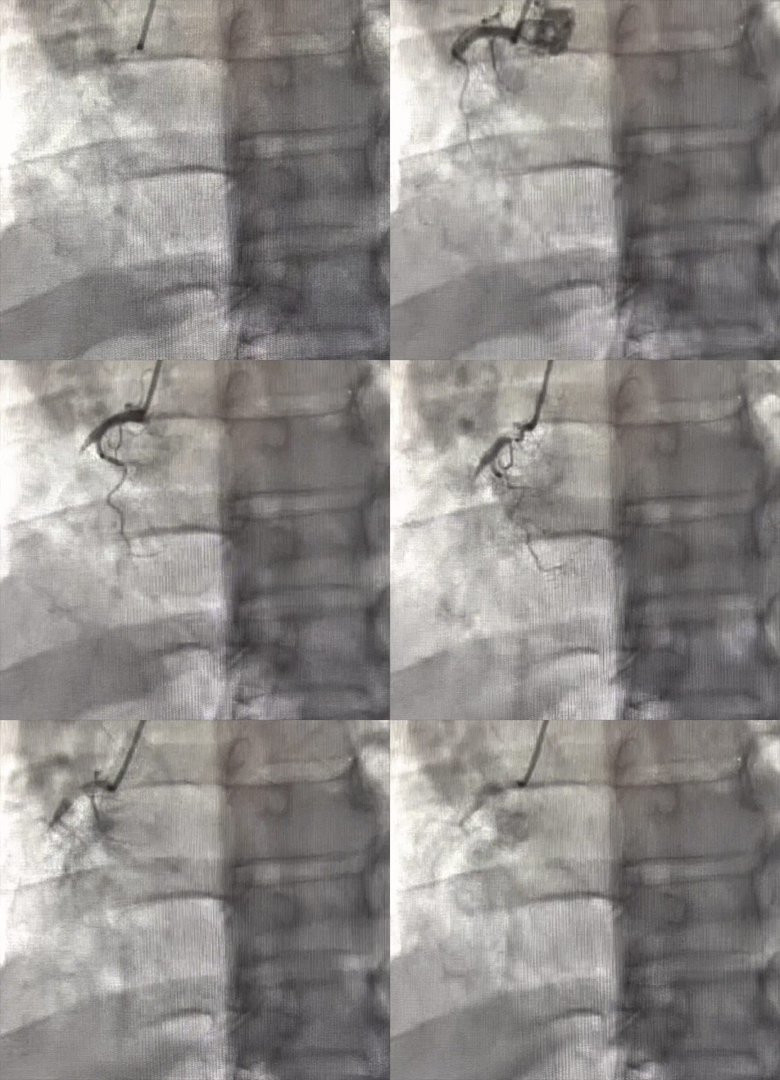
There was co-dominant circulation, with 90% stenosis of distal LCx and 100% thrombotic occlusion of proximal RCA with TIMI Flow 0. DES was deployed at 9 AM RCA with plan for staged intervention to LCx the next day.
TTE post cath showed preserved EF of 55-60% and hypokinesis of the basal to mid inferior wall of LV. There was no repeat EKG or troponin after intervention. Patient was discharged with diagnosis of NSTEMI.
Frick: If there had been a repeat troponin, it would likely have been very high (like this case where troponin stayed in the hundreds before skyrocketing to 37,000 ng/L after PCI). In the case of TIMI 0 occlusion, troponin gets "trapped" in the ischemic myocardium.
Learning points:
Prior to looking at EKG, pretest probability trumps everything. Positive QOH interpretation means very little when applied to a patient without chest pain. Similarly, QOH negativity is not completely reassuring if the pre-test probability is very high as in this case.
Ischemic heart disease is more prevalent in South Asian decents. According to a circulation review article, South Asians are at a higher rate of ASCVD, higher burden with higher.
Smith: by waiting for PCI in this "Transient OMI," the patient lost a lot of myocardium. Do not Wait!!
MY Comment, by KEN GRAUER, MD (6/10/2025):
- In a patient who had CP severe enough to prompt a visit to the ED — but whose CP had significantly decreased by the time today's initial ECG was recorded — I found ECG #1 diagnostic of acute infero-postero OMI.
- The rhythm is sinus.
- The 3 leads that immediately caught my "eye" — are lead III, lead aVL, and lead V2 (within the RED rectangles).
- Lead III immediately raises suspicion. We see a very deep (albeit narrow) Q wave + ST segment coving + terminal T wave inversion (BLUE arrow). There really is not clear ST elevation in the other 2 inferior leads (leads II and aVF) — so by itself, lead III is not definitive. But the mirror-image opposite ST-T wave picture that we see in lead aVL (ie, subtle-but-real ST depression with terminal T wave positivity) in this patient who had new CP that has now decreased — indicates acute inferior OMI until proven otherwise (See My Comment at the bottom of the page in the October 6, 2018 post and in the May 10, 2025 post, among many others — regarding clinical significance of the "magic" mirror-image opposite relationship between leads III and aVL).
- Confirmation that ECG #1 is diagnostic of acute inferior OMI — is forthcoming by clear indication of associated posterior OMI (given the very common blood supply of both inferior and posterior LV walls). As we often emphasize, there is normally slight, gently upsloping ST elevation in both leads V2 and V3. When this is missing (as the RED and BLUE arrows in these leads highlight) in a patient with suspected inferior OMI — this strongly suggests infero-postero OMI until proven otherwise.
- Reduction in CP, in association with the modest but-definitely-present ST-T wave abnormalities that we see in leads III, aVL and V2,V3 — suggest that the "culprit" vessel (probably the RCA) — has spontaneously reopened.
- But as we so often emphasize — What spontaneously reopens, may just as easily at any moment spontaneously reclose. Therefore, optimal management is not to delay cath until the next morning — but instead entails definitive treatment by prompt cath with PCI.
-USE.png) |
| Figure-1: The 1st ECG shown in today's case. |


No comments:
Post a Comment
DEAR READER: I have loved receiving your comments, but I am no longer able to moderate them. Since the vast majority are SPAM, I need to moderate them all. Therefore, comments will rarely be published any more. So Sorry.