Written by Willy Frick
At first glance there seems to be a lot of variation happening, but by focusing on one part of the ECG at a time we can make sense of it. Ignore the first QRS complex and look at beats 2 through 5. I have labeled them below for ease of reference:
We see P waves which are upright in leads I, II, and aVF, and upside down in aVR. This is consistent with sinus rhythm. (Reminder that sinus rhythm is not determined by "a P before every QRS and a QRS after every P.") What happens afterward? R waves 6 through 9 have no preceding P waves and are suspiciously regularly spaced. If we set our calipers between R waves 6 and 7 and march this out in either direction, we see the following:
Specifically, there are four regularly spaced, wide complexes not preceded by P waves. In general, this is consistent either with ventricular pacing or ventricular escape rhythm. Escape rhythms are generally extremely regular (unlike sinus rhythm which exhibits some natural variation in cycle length). This patient does not have a pacemaker, so this must be a ventricular escape rhythm.
Afterward, we see resumption of normal sinus rhythm in beats 10 through 12. So why does the sinus node apparently stop pacing for a few seconds in the middle of the ECG?
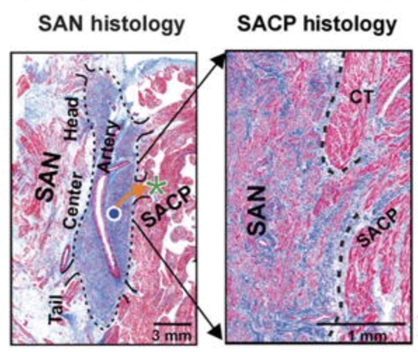
Answer: The sinus node is pacing the whole time, but exit block prevents it from depolarizing the surrounding atrium. To explain further, the sinus node (AKA sinoatrial node) is electrically insulated from the surrounding atrial myocardium which probably serves to protect automaticity from hyperpolarization due to atrial myocytes [Csepe]. Depolarization wavefronts exit the sinus node through one or several sinoatrial conduction pathways (SACP) as seen in the histology section below taken from Li et al.
It is possible for this pathway (i.e. the orange arrow) to become blocked, resulting in failure of the sinoatrial node impulse to depolarize the surrounding atrium.
How might this manifest on the surface ECG? Whenever the sinus node exit site is blocked, there would be an absent P wave. Returning to our rhythm strip below:
We can set our calipers on the P waves in beats 2 and 3 and march them out. I have added labels for ease of reference:
- First degree SA exit block is defined by prolonged SA conduction time and cannot be diagnosed on 12-lead ECG.
- Second degree SA exit block type I is defined by progressive prolongation in sinoatrial conduction time followed by a blocked impulse which then resets the cycle. This (counterintuitively) results in progressive P-P shortening, a dropped P wave, and the cycle repeats.
- Second degree SA exit block type II is defined by intermittent failure of the sinus to depolarize the surrounding atrium as in this post.
- Third degree SA exit block is defined by failure of all sinus impulses to depolarize the surrounding atrium. It is indistinguishable from sinus arrest on surface ECG.
Csepe, T. A., Zhao, J., Hansen, B. J., Li, N., Sul, L. V., Lim, P., Wang, Y., Simonetti, O. P., Kilic, A., Mohler, P. J., Janssen, P. M. L., & Fedorov, V. V. (2016). Human sinoatrial node structure: 3D microanatomy of sinoatrial conduction pathways. Progress in Biophysics and Molecular Biology, 120(1–3), 164–178. https://doi.org/10.1016/j.pbiomolbio.2015.12.011
Li, N., Hansen, B. J., Csepe, T. A., Zhao, J., Ignozzi, A. J., Sul, L. V., Zakharkin, S. O., Kalyanasundaram, A., Davis, J. P., Biesiadecki, B. J., Kilic, A., Janssen, P. M., Mohler, P. J., Weiss, R., Hummel, J. D., & Fedorov, V. V. (2017). Redundant and diverse intranodal pacemakers and conduction pathways protect the human sinoatrial node from failure. Science Translational Medicine, 9(400). https://doi.org/10.1126/scitranslmed.aam5607
MY Comment, by KEN GRAUER, MD (8/14/2024):
- The KEY to interpreting many complex arrhythmias lies with determination of what is and what is not a P wave. And then, once you identify P waves — determining whether small but definitely-present variations in P wave morphology represent "normal" P wave variation (that can sometimes be seen in shape from 1 P wave-to-the-next with sinus rhythm) — vs — atrial depolarization arising from another atrial site.
- And finally — determining whether certain P waves are forward-conducting or being conducted retrograde.
- The "good news" — is that our interpretations agree on the principal findings, namely that some sort of SA block appears to be present — and — that the result is the transient appearance of a ventricular escape rhythm at the slightly accelerated rate of ~65/minute (ie, AIVR = Accelerated IdioVentricular Rhythm). That said — our opinions differ on some of the details (ie, Dr. Frick favors a form of the more serious Type II SA block — whereas I do not believe we have enough information from this single tracing to know what kind of SA block we truly are dealing with, nor whether pacing is likely to be needed).
- The "other" good news — is that over the years, I've probably learned the most about arrhythmia interpretation from cases in which my opinion differed from that of an esteemed colleague. We learn best when we learn from each other. And sometimes we find out that both of us are equally correct — or equally wrong.
- Thus, my favorite truism about complex arrhythmias is by Rosenbaum — "Every self-respecting arrhythmia has at least 3 possible interpretations." (See the Addendum in My Comment in the September 9, 2020 post for additional illustration of this truism).
-USE.png) |
| Figure-1: I've labeled the initial ECG in today's case to illustrate my theory. |
- Type I SA Block (also known as SA Wenckebach) — is the more common form of 2nd-degree SA block. Similar to the prognostic implications of Mobitz I AV block — SA Wenckebach is not necessarily pathologic, and not necessarily associated with adverse outcome.
- In contrast — true Type II SA Exit Block (like the Mobitz II form of AV block) — is much more likely to be associated with adverse outcome and potential need for cardiac pacing. (For more re "My Take" on SA block — Please check out my Summary below in Figures-3 and -4 of today's ADDENDUM — as well as My Comments on SA block in the May 25, 2022 post and the August 30, 2023 post in Dr. Smith's ECG Blog).
- The underlying rhythm in today's tracing is sinus — as determined by the presence of upright sinus P waves in the long lead II with a constant and normal PR interval (RED arrows in Figure-1).
- In addition to the 7 narrow sinus-conducted beats — there are 5 wider beats (ie, beats #1,6,7,8 and 9). At least for the first 4 of these wider beats — there is no P wave preceding them — therefore (as per Dr. Frick) — beats #1,6,7,8 are of ventricular etiology.
- The equal R-R interval (of just under 5 large boxes in duration) between beats #6-to-7 and between #7-to-8 — indicates a rate of ~65/minute — which suggests that the wide beats in today's tracing represent AIVR (a slightly Accelerated IdioVentricular Rhythm).
- I can not explain why there is no P wave between beats #5-to-6 (the ? in Figure-1). This suggests there is some form of SA block.
- AIVR is often an "escape" rhythm that occurs when supraventricular rhythms (sinus or junctional) either slow down too much or fail completely. Thus, it is both fortunate and completely appropriate for this run of AIVR to begin with beat #6 following the SA block. Note that the R-R interval before this first ventricular beat #6 is the same as the R-R interval between beats #6-7 and between #7-8.
- Looking closer at each of the wider beats in the long lead II — the shape of these beats is different! This raises the question of WHY the shape of beats #1,6,7,8,9 changes?
- Overall — there is not much artifact in the tracing in Figure-1. As a result (since we can not attribute artifact as the reason for changing QRS morphology) — the reason for changes in QRS shape of the wider beats may be that P waves are partially hidden within them!
- I interpreted the negative deflections just after the wide QRS of beats #7 and 8 to represent retrograde P waves (YELLOW arrows in Figure-1).
- There is also a notch at the very end of the wide QRS of beat #6 — which I interpreted as a retrograde P wave with a shorter RP' interval compared to the RP' interval following beats #7 and 8 (WHITE arrow in Figure-1).
- No such notching or negative deflections follow the remaining 2 wide beats ( = beats #1 and #9). In particular — I interpreted the small, positive "hump" deforming the beginning of wide beat #9 as a sinus P wave that might be partially conducting, but which has too short of a PR interval to be able to fully conduct to the ventricles (ie, The reason wide beat #9 may be somewhat smaller than wide beats #6,7,8 — may be because beat #9 is a fusion beat).
- The only beat that I've not discussed is beat #1. That said — because we do not see what happens before beat #1 — I don't think there is much that can be said other than that this wide beat which is not preceded by any P wave is also of ventricular etiology. NOTE: If we look up from beat #1 in the long lead II rhythm strip — we see what looks like artifact distortion in simultaneously-recorded leads I and III, suggesting there is too much artifact distortion of beat #1 to say more.
- I begin my laddergram with a Question Mark over beat #1 — which, for the reasons I noted above — not much can be said beyond labeling it as a ventricular beat.
- Beats #2,3,4,5 are sinus-conducted beats. I drew them all as uniformly exiting the SA node in similar fashion, with uneventful conduction down through to the ventricles.
- The ? between beats #5 and 6 highlights failure of the next SA nodal impulse to make it out of the SA Node ( = SA block of some sort).
- There follows a 4-beat run of AIVR (beats #6,7,8,9). I chose to draw on my laddergram the continuation of fairly regular SA nodal impulses that fail to make it out of the SA Node. But as opposed to continuation of a pathologic SA Node Exit Block — Isn't it possible that there is no SA Exit Block for these next 3 P waves? Couldn't it be that retrograde conduction from the appropriate ventricular "escape" rhythm is the reason the next 3 SA nodal impulses are unable to depolarize the atria?
- I drew beat #9 as a fusion beat — with the SA Node putting out an impulse early enough to make it through the atria (the PINK arrow) — and then through the AV Node and into the ventricles before meeting (and "fusing" with) the 4th ventricular escape beat ( = beat #9).
- Note that the rate of the last 3 SA nodal impulses in this tracing (the last 3 RED arrows) speeds up a bit, resulting in these sinus P waves capturing the ventricles with sinus conduction of the last 3 beats in this tracing (occurring because the rate of these last 3 sinus P waves in now faster than the ventricular "escape" rate).
- I could have postulated a sinus "pause" occurred after beat #5 — thereby resulting in omission of the 5th SA nodal impulse ( = the 5th RED circle at the top of the SA Nodal Tier).
- I could have postulated SA Wenckebach as the cause for failure of the 5th SA nodal impulse to make it out of the SA node (in which case I would have drawn progressively slower conduction of the first 4 SA nodal impulses out of the SA Node until the 5th SA nodal impulse is dropped — in comparable fashion to the sequence of events with AV Wenckebach).
- BOTTOM Line: We simply can not determine with certainty from this single tracing what the precise mechanism for this rhythm is. But, if my interpretation that the WHITE and YELLOW arrows do represent retrograde conduction from ventricular beats #6,7,8 that goes all the way back to the atria — then it may be that no more than a single SA nodal impulse failed (or was delayed) — and the cause of today's rhythm might not adversely affect longterm outcome (and may not require a pacemaker).
-USE.png) |
| Figure-2: My proposed laddergram — illustrating my theory (See text). |
-USE.png) |
| Figure-3: Essentials of SA Block (Modified from Grauer: ACLS-2013-ePub). |
-USE.png) |
| Figure-4: Essentials of SA Block (Modified from Grauer: ACLS-2013-ePub — and from Marriott & Conover: Advanced Concepts in Arrhythmias, 1983). |


No comments:
Post a Comment
DEAR READER: I have loved receiving your comments, but I am no longer able to moderate them. Since the vast majority are SPAM, I need to moderate them all. Therefore, comments will rarely be published any more. So Sorry.