Written by Pendell Meyers
A teenager was involved in a motor vehicle collision and presented to the Emergency Department via EMS altered and potentially critically ill. He was intubated for altered mental status. Chest trauma was suspected on initial exam.
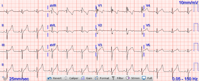
Here is his initial ECG around 1330:
The ECG shows sinus tachycardia with RBBB and LAFB, without clear additional superimposed signs of ischemia. It is very unlikely that a previously healthy teenager would have such disease of the conduction system, bringing up the possibility of blunt cardiac injury in this clinical setting.
Trauma CTs showed a "mildly displaced sternal fracture and a small retrosternal hematoma." There were no radiographic injuries noted in the head/spine/abdomen/pelvis CTs.
1445:
1520:
Troponins:
3,830 ng/L
4,098 ng/L
1,343 ng/L
Echo:
LV: normal cavity size and thickness. Systolic function is mildly reduced by visual assessment. EF 45%, with mild global hypokinesis.
RV: Cavity size normal. Systolic function normal by visual assessment only, unable to visualize well for further characterization.
 | |
|
No other significant injuries were found. The patient did well and was discharged. No cardiac MRI was done. Hopefully a repeat echocardiogram will be performed outpatient.
See our other cases of myocardial contusion and related cases (some of which have an important diagnosis OTHER THAN myocardial contusion!):
A Child with Blunt Trauma -- See how the ECG can be definite for myocardial contusion, but subtle, and what happens if you miss it.
A man in his 40s with multitrauma from motor vehicle collision
Here is some information on blunt cardiac injury from one of our prior posts:
The EKG is not generally sensitive for cardiac contusion. The right ventricle comprises the majority of the anterior heart which is most susceptible to direct injury in blunt chest trauma. Cardiac contusion can manifest on the ECG in a number of ways, including: ST segment elevation or depression, prolonged QT, new Q waves, conduction disorders such as RBBB, fascicular block, atrioventricular (AV) nodal conduction disorders (1,2, and 3 degree AV block), and arrhythmias such as sinus tachycardia, atrial and ventricular extrasystoles, atrial fibrillation, ventricular tachycardia, ventricular fibrillation, sinus bradycardia, and atrial tachycardia (Sybrandy). RBBB in blunt chest trauma seems to be indicative of several RV injury. Atrial fibrillation is also a predictor of worse outcomes in this case (Alborzi).
See these publications for more information
See these publications for more information
Overall, management for cardiac contusion is mostly supportive unless surgical complications develop, involving appropriate treatment of dysrhythmias and hemodynamic instability. Ultimately, a normal ECG and normal troponin at 4-6 hours from initial traumatic incident is highly predictive of a lack of future cardiac complications in blunt chest trauma. Between 81-95% of life-threatening ventricular dysrhythmias and acute cardiac failure occur within 24-48 hours of hospitalization. Troponins and EKGs should be trended until normalization (Sybrandy).
Delayed cardiac rupture is a potential consequence, especially if there is any ST Elevation. See this case, this case, and this case. In patient's at risk, physical activity should be limited for several months after the injury.
References
Alborzi, Z., Zangouri, V., Paydar, S., Ghahramani, Z., Shafa, M., Ziaeian, B., Radpey, M. R., Amirian, A., & Khodaei, S. (2016, April 13). Diagnosing myocardial contusion after blunt chest trauma. The journal of Tehran Heart Center. Retrieved July 2, 2022, from https://www.ncbi.nlm.nih.gov/pmc/articles/PMC5027160/
Moyé, D. M., Danielle M. Moyé From the Division of Cardiology, Dyer, A. K., Adrian K. Dyer From the Division of Cardiology, Thankavel, P. P., Poonam P. Thankavel From the Division of Cardiology, & The Data Supplement is available at http://circimaging.ahajournals.org/lookup/suppl/doi:10.1161/CIRCIMAGING.114.002857/-/DC1.Correspondence to Poonam Punjwani Thankavel. (2015, March 1). Myocardial contusion in an 8-year-old boy. Circulation: Cardiovascular Imaging. Retrieved July 2, 2022, from https://www.ahajournals.org/doi/10.1161/CIRCIMAGING.114.002857
Sybrandy, K. C., Cramer, M. J. M., & Burgersdijk, C. (2003, May). Diagnosing cardiac contusion: Old Wisdom and new insights. Heart (British Cardiac Society). Retrieved July 2, 2022, from https://www.ncbi.nlm.nih.gov/pmc/articles/PMC1767619/
===================================
MY Comment, by KEN GRAUER, MD (2/6/2024):
===================================
Today's case by Dr. Meyers provides insight with regard to sequential evolution of serial ECGs during the course of cardiac contusion.
- For clarity in Figure-1 — I've reproduced and labeled a number of findings in the initial ECG from today's case.
The Initial ECG in Today's Case:
As per Dr. Meyers — the initial ECG in today's case shows sinus tachycardia with bifascicular block ( = RBBB/LAHB). Especially in view of Dr. Meyers point that a previously healthy teenager would be unlikely to manifest such significant ECG abnormalities — I thought there were a number of additional ECG indicators of injury severity. These include:
- Marked fragmentation of the QRS complex in lead V1 — which rather than manifesting a clear triphasic rsR' complex expected when RBBB occurs in an otherwise healthy young adult — shows an all positive and slurred upright and markedly widened (to ≥0.16 second) complex in this lead (within the RED oval in Figure-1).
- Small-but-present initial Q waves are seen in 4/6 chest leads (ie, in leads V2,V3,V4,V5 — as per the RED arrows in Figure-1). Q waves in association with RBBB are usually not seen in anterior leads unless there is pulmonary hypertension or anterior infarction.
- Although ST-T wave appearance in Figure-1 is unlikely to be the result of an acute OMI in today's previously healthy teenage trauma victim — I thought at least 4/12 leads in ECG #1 manifested potentially hyperacute changes, including: i) The ST-T wave in lead V1 looks disproportionately deep, with depth of T wave inversion comparable to height of the R wave in this lead (within the BLUE rectangle in Figure-1); and, ii) The ST segment takeoff in leads V4, V5, and V6 is straightened, having lost the normal slight, gentle upsloping of chest lead ST segments (within the BLUE rectangles in these leads). The ST-T wave in leads V5,V6 looks to be disproportionately large (ie, hyperacute) given QRS amplitude in these leads.
-USE.png) |
| Figure-1: The initial ECG in today's case. |
What are the ECG Findings of Cardiac Contusion?
I've copied KEY points from My Comment in the August 6, 2022 post in Dr. Smith's ECG Blog — regarding the answer to this question.
- The ECG is less than optimally sensitive for detecting cardiac injury following blunt trauma. This is because the anterior anatomic position of the RV (Right Ventricle), and its immediate proximity to the sternum — makes the RV much more susceptible to blunt trauma injury than the LV.
- CAVEAT: Although the RV is much more susceptible to blunt trauma injury than the LV — Because of the much greater electrical mass of the LV, electrical activity (and therefore ECG abnormalities) from the much smaller and thinner RV are more difficult to detect.
To REVIEW (Sybrandy et al: Heart 89:485-489, 2003 — Alborzi et al: J The Univ Heart Ctr 11:49-54, 2016 — and Valle-Alonso et al: Rev Med Hosp Gen Méx 81:41-46, 2018) — ECG findings commonly reported in association with Cardiac Contusion include the following:
- None (ie, The ECG may be normal — such that not seeing any ECG abnormalities does not rule out the possibility of cardiac contusion).
- Sinus Tachycardia (common in any trauma patient ...).
- Other Arrhythmias (PACs, PVCs, AFib, Bradycardia and AV conduction disorders — potentially lethal VT/VFib).
- RBBB (as by far the most common conduction defect — owing to the more vulnerable anatomic location of the RV). Fascicular blocks and LBBB are less commonly seen.
- Signs of Myocardial Injury (ie, Q waves, ST elevation and/or depression — with these findings suggesting LV involvement).
- QTc prolongation.
- NOTE: Prediction of cardiac contusion "severity" on the basis of cardiac arrhythmias and ECG findings — is an imperfect science.
Additional KEY Points:
Despite the predominance for RV (rather than LV) injury — use of a right-sided V4R lead has not been shown to be helpful compared to use of a standard 12-lead ECG for detecting ECG abnormalities.
- In addition to ECG abnormalities related to the blunt trauma of cardiac contusion itself — Keep in mind the possibility of other forms of cardiac injury in these patients (ie, valvular injury, aortic dissection, septal rupture) — as well as the possibility of a primary cardiac event (ie, acute MI may have been the cause of an accident that led up to the trauma).
- ECG abnormalities may be delayed — so repeating the ECG if the 1st tracing is normal is appropriate when concerned about severe traumatic injury.
- That said — IF troponin is normal at 4-6 hours and IF the ECG is normal — then the risk of cardiac complications is extremely low.
Final COMMENT: While the literature does not provide us with specific ECG criteria for assessing severity of cardiac contusion — today's case does provide insight as to how clinical correlation with serial ECGs can confirm that the patient is recovering.
- I've noted above the plethora of distinct ECG abnormalities associated with today's initial tracing. As per Dr. Meyers — all ECG abnormalities normalized over the course of 5 sequential tracings.
- As abnormal as the initial ECG was in Figure-1 — the final ECG in today's case was a normal tracing, which correlated with dramatic improvement in the patient's condition, resulting in ultimate discharge home from the hospital.


No comments:
Post a Comment
DEAR READER: I have loved receiving your comments, but I am no longer able to moderate them. Since the vast majority are SPAM, I need to moderate them all. Therefore, comments will rarely be published any more. So Sorry.