Occlusion myocardial infarction is a clinical diagnosis
Written by Willy Frick (@Willyhfrick). Willy is a cardiology fellow with a keen interest in the ECG in OMI.
A woman in her late 70s presented with left arm pain. The arm pain started the day prior when she was at the dentist's office for a root canal. Her systolic blood pressure at the dentist was over 200 mm Hg. She was given nitroglycerin which improved her blood pressure, and she completed the procedure. Her arm pain abated. The pain returned that evening and woke her from sleep. She eventually fell back asleep, and woke up feeling normal the next day (the day of presentation). After dinner the day of presentation, she had left neck and elbow pain which she described as dull, achy, and worse with exertion. She contacted her neighbor, a nurse, for help. The neighbor recorded a systolic blood pressure again above 200 mm Hg and advised her to come to the ED to address her symptoms.
The patient presented to triage at around 10 PM. Triage documented a complaint of left shoulder pain. The patient said, "I just don't feel good." If an immediate EKG was obtained, it was not saved in the medical record. The patient had a first hsTnI drawn at 10:30 PM, which resulted at 202 ng/L (ref. <14). The first available EKG was recorded just after midnight, presumably around the time the result of the troponin came to clinical attention. It is not known what symptoms she was experiencing at this time, but it is reasonable to assume she had persistence of her presenting symptoms, especially as she had received no treatment thus far.
EKG1
The Queen of Hearts sees no OMI with high confidence. She seems a little concerned about some very subtle depression in the high lateral leads, but otherwise sees a pretty benign tracing. Recall from this post referencing this study that "reciprocal STD in aVL is highly sensitive for inferior OMI (far better than STEMI criteria) and excludes pericarditis, but is not specific for OMI." Is there anything else on the tracing to corroborate inferior OMI?
Smith: that subtle inferior STE with subtle reciprocal STD in I and aVL is highly suspicious for inferior OMI, but with the T-wave inverted in III, probably reperfusing OMI. When I see this, I always look at V2 for any evidence of posterior OMI (STD, or loss or inversion of T-wave, or downsloping ST segment: there is a negative T-wave), and V6 for any STE (normal). So I would be worried about inferior OMI. Moreover, the patient has ongoing symptoms and has an unexplained elevated troponin, so she is having an MI and the only question is whether it is type 1 or type 2 due to hypertension.
Smith again: Whenever someone sends me an ECG for determination of OMI or Not OMI, I say: "Any patient with OMI can have an ECG that does not show OMI. A patient with OMI can have a totally normal ECG!" This is especially true for circumflex occlusion. See this case: Persistent Chest Pain, an Elevated Troponin, and a Normal ECG. At midnight.
Case continued
She was loaded with aspirin 325 mg, and repeat troponin drawn around the time of EKG 1 resulted at 267 ng/L.
What would you do at this time with this information?
About an hour later, the patient was given labetalol 20 mg IV and started on continuous heparin infusion. A third troponin was drawn, which eventually resulted at 450 ng/L, a clear dynamic rise. Cardiology was consulted, and repeat EKG was obtained at around 2:30 AM. It is not known what the patient's symptoms were at this time.
EKG2
The Queen of Hearts again sees no OMI with high confidence. The subtle ST depressions in the high lateral leads have resolved. You may notice a few other very subtle differences, but it is hard to know what to make of them. There T waves in I/aVL are little bit more upright and symmetric, and the T waves in V1-2 which were previously inverted are now essentially flat. Is this normal variation in baseline EKG or dynamic ischemic change? The presence of rising troponin and anginal symptoms certainly argues for the latter, but the changes are extremely subtle. The resolution of high lateral STD and appearance of more symmetric, upright T waves raises the possibility of reperfusing inferior OMI. These changes are shown below side-by-side.
In this version 1, the Queen of Hearts does not compare serial ECGs. She cannot see this change!
Shortly after this EKG was obtained, the patient received morphine 2 mg IV with improvement in her symptoms. By the time she was seen by cardiology her pain was better, but it is not clear if it was resolved. The cardiology note indicates a diagnosis of suspected type 1 NSTEMI and a plan for same day cardiac catheterization.
A few notes:
- Morphine treats ALL forms of pain. Patients will feel better after receiving morphine. But pain is an important signal in MI and informs the clinician of the urgency. You would not delay surgery for a ruptured appendix because pain resolved with morphine. For the same reason, you should not delay coronary angiography because pain resolves with morphine. This is different from nitroglycerin which produces vasodilation and can improve by pain improving myocardial perfusion.
- Both the outdated 2014 AHA/ACC guidelines and the updated 2023 ESC guidelines recommend immediate invasive management of patients with uncontrolled chest pain. Remember that OMI is a clinical diagnosis which usually produces diagnostic EKG changes, but not always.
- Recall that RIDDLE-NSTEMI enrolled patients similar to this, and showed a 68% reduction in death or MI at one month in patients managed with immediate invasive (< 2 hours) vs delayed invasive (2-72 hours) treatment. The results were sustained at one year of follow up.
Smith: As Willy states, ACS with persistent symptoms is a guideline recommended indication for <2 hour angio (both ACC/AHA and ESC). The ESC states that patients with suspected ACS should go to the cath lab in <2 hours "regardless of ECG or biomarker evidence of MI!!" Unfortunately, the only study of compliance with this high-risk ACS guideline showed that this guideline is followed only 6% of the time. We have shown that morphine is associated with worse outcomes (see learning points below) and have published many blog posts about it, such as this one: Another myocardial wall is sacrificed at the altar of the STEMI/NonSTEMI mass delusion (and Opiate pain relief).
The case continues...
Repeat troponin drawn four hours after the last increased from 450 ng/L to 1366 ng/L. A third EKG was performed around 8 AM.
EKG 3
The Queen of Hearts sees no OMI with high confidence. It is hard to see much difference between EKG 2 and 3. The T waves in I/aVL are slightly taller, which could be evidence of persistent reperfusion of an inferior OMI. Four hours later around noon, the patient's symptoms returned and were severe. A fourth EKG was obtained.
EKG 4
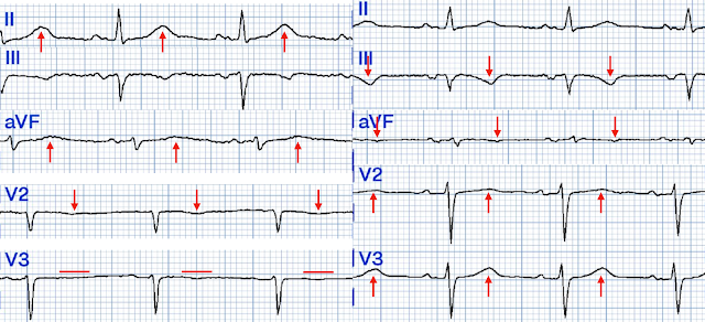
The Queen of Hearts sees no OMI with high confidence. Notice that as the patient's symptoms are worsening, the area under the curve of the T wave in II is suddenly much larger (possibly hyperacute), and the previously flat T waves in V2-3 are now subtly inverted. It is so subtle, it is not even clearly visible in every beat. This could be consistent with re-occlusion of an inferoposterior MI. This is shown below.
A few hours later, the patient underwent coronary angiography, which showed complete occlusion of her mid left circumflex artery.
After returning from lab repeat troponin was 20,380 ng/L, and later that evening it peaked at 29,571 ng/L before trending down. The patient suffered a large infarct. Her contrast enhanced echocardiogram is shown below in the parasternal short axis view. Notice that there is much less thickening between the red lines, in the inferolateral wall (which is another way of saying posterior). It is severely hypokinetic or even akinetic.
One final EKG was obtained the next morning, shown below.
EKG 5
Especially now that we know the diagnosis, we are able to appreciate the following changes.
- The T waves in II are no longer hyperacute
- The T waves in III are more deeply inverted, consistent with reperfusion
- The T waves in aVF are subtly inverted, consistent with reperfusion
- The T waves in V2-3 have gone from inverted/flat to upright, reciprocal to inverted posterior reperfusion T waves
Learning Points
- OMI is a clinical diagnosis. There are usually diagnostic EKG findings but not always. EKG findings can be extremely subtle, to the point where even a highly trained, highly accurate artificial intelligence can miss them.
- You do not need to be better than the Queen of Hearts at EKG to understand that refractory chest pain NEEDS CATH NOW. Medicine constantly reminds us of the paramount importance of history taking, a requisite human skill to work in concert with AI.
- In the context of concerning chest pain or chest pain equivalent, a slowly rising troponin can lull the clinician into a false sense of security. This patient had a few troponin measurements that were elevated, but not dramatically so. In the end, she suffered a large infarct that could have been prevented by earlier intervention. If you wait until the troponin is "really convincing," you have already lost a lot of myocardium. This would be like seeing your basement flood, and patiently observing until your living room is underwater as confirmation of a problem before finally activating the sump pump.
- Morphine neutralizes pain. But pain is a critical signal for urgency in the context of acute coronary syndrome. If you are giving morphine for pain that is refractory to nitrates, the patient should be on the way to cath lab!
- Sometimes reciprocal changes are more obvious than the primary change. In this case, perhaps the easiest finding to appreciate was the STD in I and aVL in EKG 1. Remember that STD in aVL is highly sensitive for inferior OMI, although it is not specific.
Learning Points references:
Amsterdam, E. A., Wenger, N. K., Brindis, R. G., Casey, D. E., Ganiats, T. G., Holmes, D. R., Jaffe, A. S., Jneid, H., Kelly, R. F., Kontos, M. C., Levine, G. N., Liebson, P. R., Mukherjee, D., Peterson, E. D., Sabatine, M. S., Smalling, R. W., & Zieman, S. J. (2014). 2014 AHA/ACC guideline for the management of patients with non–ST-elevation acute coronary syndromes. Circulation, 130(25). https://doi.org/10.1161/cir.0000000000000134
Bischof, J. E., Worrall, C., Thompson, P., Marti, D., & Smith, S. W. (2016). St depression in lead AVL differentiates inferior st-elevation myocardial infarction from pericarditis. The American Journal of Emergency Medicine, 34(2), 149–154. https://doi.org/10.1016/j.ajem.2015.09.035
Byrne, R. A., Rossello, X., Coughlan, J. J., Barbato, E., Berry, C., Chieffo, A., Claeys, M. J., Dan, G.-A., Dweck, M. R., Galbraith, M., Gilard, M., Hinterbuchner, L., Jankowska, E. A., Jüni, P., Kimura, T., Kunadian, V., Leosdottir, M., Lorusso, R., Pedretti, R. F., … Zeppenfeld, K. (2023). 2023 ESC guidelines for the management of acute coronary syndromes. European Heart Journal, 44(38), 3720–3826. https://doi.org/10.1093/eurheartj/ehad191
Milosevic, A., Vasiljevic-Pokrajcic, Z., Milasinovic, D., Marinkovic, J., Vukcevic, V., Stefanovic, B., Asanin, M., Dikic, M., Stankovic, S., & Stankovic, G. (2016). Immediate versus delayed invasive intervention for non-stemi patients. JACC: Cardiovascular Interventions, 9(6), 541–549. https://doi.org/10.1016/j.jcin.2015.11.018
Opiates are associated with worse outcomes in Myocardial Infarction.
See this case: A man his 50s with chest pain. What happens when you treat with morphine rather than with reperfusion?
----See this study showing an association between morphine and mortality in ACS:
Use of Morphine in ACS is independently associated with mortality, at odds ratio of 1.4. Meine TJ, Roe M, Chen A, Patel M, Washam J, Ohman E, Peacock W, Pollack C, Gibler W, Peterson E. Association of intravenous morphine use and outcomes in acute coronary syndromes: Results from the CRUSADE Quality Improvement Initiative. Am Heart J. 2005;149:1043–1049.
And Another that we wrote:
65 (23.9%) patients were found to have STEMI(-) occlusion myocardial infarction (OMI) at the time of cardiac catheterization. The 45 patients with STEMI(-) OMI without pre-cath opioids had a door-to-balloon time of 75 minutes, vs. 684 minutes for the 25 STEMI(-) OMI with pre-cath opioids.
High Risk ACS guidelines are only followed in 6% of patiients:
Lupu L, Taha L, Banai A, Shmueli H, Borohovitz A, Matetzky S, Gabarin M, Shuvy M, Beigel R, Orvin K, Minha S ’ar, Shacham Y, Banai S, Glikson M, Asher E. Immediate and early percutaneous coronary intervention in very high-risk and high-risk non-ST segment elevation myocardial infarction patients. Clin Cardiol [Internet]. 2022;Available from: https://onlinelibrary.wiley.com/doi/10.1002/clc.23781
MY Comment, by KEN GRAUER, MD (12/11/2023):
- Today's case illustrates how the omission of correlating the presence and relative severity of symptoms with each ECG — can easily explain the failure to recognize ongoing OMIs in a timely fashion.
- It is EASY to correlate the presence and severity of symptoms with each and every ECG during the course of a patient's event. Simply write down in the chart (and/or on each ECG) — IF CP was or was not present at the time a given tracing was recorded — and, IF present — the relative severity of such CP on a scale from 1-to-10. Unfortunately, this was not done in today's case.
- I suspect the principal reason why the above information is so commonly omitted in the cases I review — is a lack of appreciation of the pathophysiology of acute OMI. Acute coronary occlusion typically results in CP and acute ST-T wave changes (ST elevation and/or other acute ECG findings).
- Acute reperfusion of the "culprit" vessel — is typically accompanied by normalization of abnormal ECG findings in association with reduction or resolution of CP.
- Spontaneous reperfusion of the "culprit" artery is not uncommon. Clinically this is recognized in the same way reperfusion following PCI or thrombolytic therapy is recognized — ie, by reduction in CP and improvement of ST-T wave abnormalities.
- BUT — unless notation is made of the clinical course of symptom severity in association with each ECG that is done — spontaneous reperfusion will all-too-often be overlooked. We have illustrated this concept numerous times in prior posts in Dr. Smith's ECG Blog (See My Comment in the July 12, 2023 post — the May 18, 2023 post — and the April 9, 2023 post, among many others).
- BOTTOM LINE: Awareness and application of the above concept of spontaneous reperfusion resulting in "pseudo-normalization" of the ECG, with no more than nonspecific ST-T wave abnormalities — could have (should have) prompted a much earlier diagnosis of acute OMI in today's patient, probably as soon as the 1st Troponin came back with significant elevation.


No comments:
Post a Comment
DEAR READER: I have loved receiving your comments, but I am no longer able to moderate them. Since the vast majority are SPAM, I need to moderate them all. Therefore, comments will rarely be published any more. So Sorry.