The patient is a 70-something female with DMII, HTN and an extensive prior history of coronary artery disease and myocardial infarctions. She's had multiple PCI procedures. She also has sick sinus syndrome (SSS) and intermittent high grade AV block for which she had a dual chamber pacemaker implanted. On the day of presentation she complained of typical chest pain, and stated it feels like prior MI.
Just from the medical hx and clinical presentation this patient is very likely to be having an MI. The question is, does she need to go urgently to the cath lab or can she wait. Below is the presentation ECG
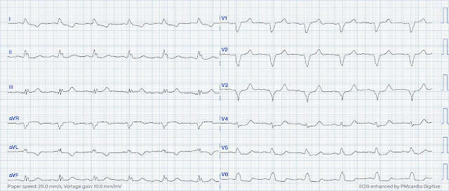
At the time of recording the ECG the patient was complaining of chest discomfort 4/10. What is your interpretation?
ECG#1
 |
It would help knowing more about the PM and specifically pacing statistics. In this case there had been 100% ventricular pacing since 2016. This makes ventricular pacing almost certain in this ECG even though 100% ventricular pacing in the statistics/memory of the PM doesn’t preclude intermittent AV conduction.
Atrial pacing spikes are not discernable. So this could be sinus rhythm with ventricular pacing or dual chamber pacing. There is likely ventricular pacing in the septum of the RV since the QRS axis does not fit with RV apical lead placement.
Regardless of the cause of the wide complexes, and whether you identified the likely pacemaker rhythm, you will still have to continue with your assessment of QRS complexes, ST-segments and T waves in this patient with new onset chest pain. Below the ECG image quality is enhanced using the PM Cardio App. What do you think?
ECG#1
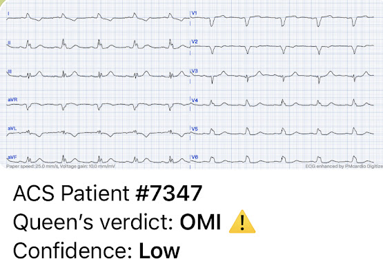
The ECG on file (ECG #2) shows findings very similar to the ECG at presentation; however, inferior T-waves are not quite as large AND the ST-T-wave in V2 appears non-ischemic (confirming that there is a real change in V2). This is very suggestive that ECG 1 does represent new OMI.
However, in ECG #2, there is pathological ST depressions in the precordial leads ALSO suggesting OMI or subendocardial ischemia. Most noticeably the ST depression in lead V4 compared to the amplitude of the preceding QRS is massive. There again is inappropriate (almost) isoelectric ST segment in V2 with a hint of ST dep V3. There might be some more absolute ST depression in V5 compared to V4, but proportionally the ST segment shift is greatest in V4.
Thus: Can this ECG be used for comparison?
You will need to assess under what circumstances the ECG on file (ECG#2) was recorded before you can use it as baseline for comparison. In fact, this patient had an extensive hx of prior MIs. The ECG (ECG#2) on file that was given you was obtained three months prior. At that visit the patient was found to have an in-stent RCA occlusion. Thus, unless you know that, you might think that there is not a significant change, since the inferior T-waves in both ECGs appear hyperacute.
Based on the ECG findings in todays case (ECG#1) the patient was suspected of having an acute OMI in the setting of a wide complex QRS. The patient was referred emergently to the cath lab, and again there was an in-stent RCA occlusion. Troponin I peaked at 18.323ng/L.
The patient's baseline ECG (ECG#3) (recorded at a time without ischemic symptoms) is shown below. As you can see, the hyperacuity of inferior wall T waves was not present at that time so this is a new finding that occurred since this non-ischemic baseline. ST segment in lead V2 has slight ST elevation as expected. Lead V3 from the two first ECG should probably not be compared to lead V3 in the baseline ECG as there is some difference in R wave amplitude likely explained by different electrode placement.
ECG#3 - no ischemia
- Make sure that when comparing to an old ECG, that it is truly baseline ECG (No ischemia)
- Isoelectric ST segments may be indicative of OMI in the setting of wide comples QRS
- Sometimes colleagues are unfamiliar with subtle ischemic changes and then you need to advocate for your patients
- Pacemaker spikes can be difficult to visualize on the ECG.
- With the PM Cardio App image quality generally is improved, however, in this case it was more difficult to see pacing spikes.
- Surprisingly — It can sometimes be extremely difficult to tell IF a rhythm is paced or not paced. Obviously — when pacer spikes are clearly seen, this distinction is easy. But pacer spikes are not always seen on an ECG, even when we are looking for them because: i) The wrong filter settings for optimal pacer spike visualization are often used (See this article by Sun et al — Chin Med J: 13295): 534-541, 2019); ii) There may be baseline artifact indistinguishable from small amplitude pacer spikes; and/or — iii) The location of pacing wires may be such that the amplitude of pacing spikes is very small.
- In practice — We often do not know the particular pacemaker specifications for a given device (ie, What is the default heart rate? — or what is the programmed PR interval after paced or spontaneous P waves before ventricular pacing is triggered?). This is the source of additional reasons why it is sometimes so difficult to tell IF a rhythm is paced — and/or — IF a pacemaker is appropriately functioning.
- The only way to know IF ST-T wave changes that we see on a paced tracing are "new" vs longterm, as the result of ventricular pacing — is IF we have a prior (baseline) paced ECG on the patient.
- And as shown above by Dr. Nossen — even when we do find a previous ECG on our patient — We need to know the circumstances at the time that "baseline" tracing was recorded (ie, Since in today's case — the first "baseline" tracing found was not truly a baseline, since it was recorded in association with a previous acute event).
- Added to the above — there is always the possibility that pacing wires may move — in which case, QRS morphology (and therefore also ST-T wave morphology) may change, thus invalidating what we thought was a baseline tracing.
- As per Dr. Nossen in today's case — Regardless of whether some or all QRS complexes in today's tracings are spontaneous — or paced — or intermittently paced — or consist of fusion beats produced by near simultaneous pacing with sinus-conducted P waves — Dr. Nossen describes in detail the inappropriate ST segment flattening, as well as hyperacute-looking T waves — that would be abnormal regardless of whether QRS complexes were paced or spontaneous.
- PLUS — Today's patient is an older woman with known severe coronary disease who presented with new chest pain. While fully aware that ECG assessment will not be perfectly predictable in this setting — there should be no doubt that prompt cath was needed in today's case!


No comments:
Post a Comment
DEAR READER: I have loved receiving your comments, but I am no longer able to moderate them. Since the vast majority are SPAM, I need to moderate them all. Therefore, comments will rarely be published any more. So Sorry.