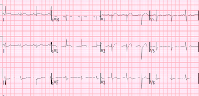
A male in his 30's complained of sudden severe substernal chest pain. He was rushed to the critical care area where they recorded this ECG:
The cath lab was activated, and just before going to the cath lab (19 minutes after the first ECG), this ECG was recorded:
I have often wondered if de Winter's T-waves really are due to complete occlusion, or to severe, subtotal occlusion. Perhaps they indicate an open artery with minimal flow and severe subendocardial ischemia, but not total subepicardial ischemia.
Since ACS is so dynamic, with thrombi forming and lysing continuously, and because the ECG and angiogram are rarely simultaneous, it is probable that de Winter's T-waves are recorded in a window when the artery is barely open.
At cath, there was a 99% lesion with some flow.
Here is the first post cath ECG, shortly after opening.
Here is the ECG the next day:
de Winter's waves
de Winter et al. (Heart 2009;95:1701–1706, http://dare.uva.nl/document/214264) published on 1890 patients who had LAD occlusion. They found these "de Winter's waves" (ST depression in V1-V6 with upsloping ST depression and a hyperacute T-wave) in 2% of these patients, and stated they were "persistent."
A quote: "We have observed this pattern as a static ECG pattern lasting from the time of first medical contact until the recording of the pre-procedural ECG and lasting until angiographic establishment of an occluded LAD artery (that is, approximately 60 minutes)"
They gave as an example a patient with chest pain of 91 minutes who had this pattern. He had an ECG 71 minutes later and the pattern continued. Then, they say: "The coronary angiogram performed shortly after this registration revealed a fresh occlusion of the proximal LAD artery." It is in my view that it is very likely that the artery was barely open while the de Winter's waves were present, and closed off entirely by the time of the angiogram which was done "shortly after" the ECG.
My interpretation of this data
de Winter's waves are probably due to severe subendocardial ischemia, with some epicardial ischemia (enough to result in hyperacute T-waves, but not enough for ST elevation.
This is somewhat academic, because these patients clearly need reperfusion whatever the exact thrombotic state of the artery.
The cath lab was activated, and just before going to the cath lab (19 minutes after the first ECG), this ECG was recorded:
 | |
|
I have often wondered if de Winter's T-waves really are due to complete occlusion, or to severe, subtotal occlusion. Perhaps they indicate an open artery with minimal flow and severe subendocardial ischemia, but not total subepicardial ischemia.
Since ACS is so dynamic, with thrombi forming and lysing continuously, and because the ECG and angiogram are rarely simultaneous, it is probable that de Winter's T-waves are recorded in a window when the artery is barely open.
At cath, there was a 99% lesion with some flow.
Here is the first post cath ECG, shortly after opening.
 |
| (QTc = 383). Now there are very subtle Wellens waves |
Here is the ECG the next day:
 |
| QTc 440. Well developed Wellens' waves (reperfusion T-waves after opening of LAD occlusion) |
de Winter's waves
de Winter et al. (Heart 2009;95:1701–1706, http://dare.uva.nl/document/214264) published on 1890 patients who had LAD occlusion. They found these "de Winter's waves" (ST depression in V1-V6 with upsloping ST depression and a hyperacute T-wave) in 2% of these patients, and stated they were "persistent."
A quote: "We have observed this pattern as a static ECG pattern lasting from the time of first medical contact until the recording of the pre-procedural ECG and lasting until angiographic establishment of an occluded LAD artery (that is, approximately 60 minutes)"
They gave as an example a patient with chest pain of 91 minutes who had this pattern. He had an ECG 71 minutes later and the pattern continued. Then, they say: "The coronary angiogram performed shortly after this registration revealed a fresh occlusion of the proximal LAD artery." It is in my view that it is very likely that the artery was barely open while the de Winter's waves were present, and closed off entirely by the time of the angiogram which was done "shortly after" the ECG.
My interpretation of this data
de Winter's waves are probably due to severe subendocardial ischemia, with some epicardial ischemia (enough to result in hyperacute T-waves, but not enough for ST elevation.
This is somewhat academic, because these patients clearly need reperfusion whatever the exact thrombotic state of the artery.


There's one question I've been meaning to ask for a while. In the classic ECG in STEMI there is the stage with negative T-waves, even in the untreated MI (closed artery that stays closed, right?). So how do you differentiate that from reperfusion T-waves?
ReplyDeleteAna,
Delete2 ways:
1) when a transmural MI is completed, there is indeed T-wave inversion but it is shallow. The depth correlates with the amount of viable myocardium remaining
2) when complete transmural MI, expect Q-waves, especially QS-waves
Steve
So is the depth of the T-wave directly proportional to the viable myocardium remaining? I'm just thinking that there are deep T-waves in aneurysms, where there is no viability anymore. Or do they get deeper in a late stage?
DeleteThe T-waves are rarely deeply inverted in aneurysm. There are exceptions, I am sure. But I haven't seen any. Usually well developed Q-waves with shallow T-wave inversion. On the other hand, well developed STEMI that reperfuses before complete transmural infarction still has a significatn amount of viable myocardium and can have very deeply inverted T-waves.
DeleteThank you for clarifying this for me!
DeleteDeWinter's wave- explanation is very good.Thanks.
ReplyDeleteDr.Chid
NICE post Steve! Interesting how over the past several years since I've been looking for DeWinter T waves - I've seen this phenomenon more and more in a variety of "partial forms" - each of which I imagine shows some state of high-grade narrowing but incomplete occlusion. I like your theory on the operative physiology! THANKS for posting - :)
ReplyDeletewould you thrombolyse someone with DeWinter T-waves? Will it provide benefit like PCI will?
ReplyDeleteJoseph,
DeleteI would definitely thrombolyse a patient with de Winter's T-waves. Believe it or not, the data against thrombolysing ST depression is nowhere near convincing. It was done in 4 studies: ISIS-2, GISSI-1, LATE, and TIMI IIIB. Numbers were small, time to thrombolysis was long (in TIMI IIIB, the most imporant study against ST depression, mean time to tPA was 9 hours. In GISSI, only 2 leads with ST depression of 1 mm were required. So the data on thrombolysis of severely stenotic but open thrombotic lesions, manifesting as ST depression, does not exist.
Steve Smith
Steve...
ReplyDeleteI just encountered your post here a year later (June 2015). I think you really hit the nail on the head when you relate the De Winter T waves to subendocardial ischemia. They certainly fit the morphology of regional subendocardial ischemia. Regional subendocardial ischemia is characterized by ST depression with upright T waves. It is not so generalized as circumferential subendocardial ischemia and is typically due to a partial occlusion of a major vessel or complete occlusion of a less extensive branch. The first stage of ischemia - even for subendocardial ischemia - is the appearance of hyperacute T waves. However, the ST depression of subendocardial ischemia is certainly not limited to an upward sloping ST segment.
Thanks for the great posts!
Jerry W. Jones, MD FACEP FAAEM
Jerry,
DeleteThanks for the feedback. Great comments.
Steve
So the wellens waves indicate complete reperfusion while de winters indicate only small reperfusion ??
ReplyDeleteIam right or not?
This is what I believe, and have evidence for, but it is hard to prove
DeleteHi Smith, why the second ECG just before cath is showing changes only in anterior leads while no upslopping st depression is noted in high lateral leads, another question what is reason behind doing ECG just before Cath, i mean they have the confirmed diagnosis and cath lab is already activated?
ReplyDeletethanks
1) most ischemia is gone from the lateral leads, as also seen in V5, V6.
Delete2) Waiting for cath team to arrive