This is a repost of a repost of a case from 2014. The original Case is here, and has many more details if you want to read those.
At the time, I was not nearly as good at appreciating subtle OMI as I am now.
The patient presented with syncope and trapezius pain, and some dizziness and vague left sided chest discomfort.
Here is the ED ECG:
A pediatric MD who was learning bedside echo and had only performed 20 cardiac ultrasounds in his life was sent in to practice ultrasound on this patient, not expecting to find anything, or even to be able to find anything. He recorded this echo from the apical 4-chamber view:
Echo 1 done by newbie from Stephen Smith on Vimeo.
This 4 chamber view was recorded by an emergency echo expert:
Echo 13 from Stephen Smith on Vimeo.
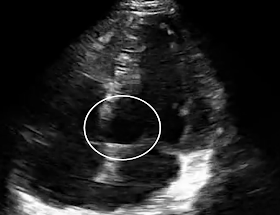
Here is a still frame with a circle that shows the area of wall motion abnormality:
 |
| This is taken in systole. The base of the septum (at the left of the circle) is not contracting as is the more apical portions of the septum. |
Here is a two chamber view (LV and LA only, with the probe turned 90 degrees from the apical 4-chamber) which shows the infero-posterior wall on the left side of the image.
2 chamber view from Stephen Smith on Vimeo.
Here is a still frame in systole showing the wall motion abnormality at the base of the heart on the left side of the picture:
Here is a map of the echocardiographic wall motion abnormalities:
 |
| Reproduced with permission of Robert F. Reardon, from Ma, Mateer, Reardon, and Joing, Editors. Emergency Ultrasound. McGraw Hill 2014. The chart shows the coronary supply of the segments of the heart as seen on various views. The circle shows the area of wall motion abnormality in this patient's apical 4 chamber view. The lavendar on the "long axis" 2 chamber view correlates with this patient's 2 chamber view. Both are consistent with the RCA involvement, and thus correlates perfectly with the ECG. |
Here are the subsequent events:
This shows how even a novice may be a able to see wall motion abnormalities that, especially if they correlate with the subtle ECG findings, may help to make the diagnosis of ACS.
Warning:
I would warn that wall motion abnormalities may be very difficult to see, and that there are also false positives.
It frequently requires echo contrast, and an expert echocardiographer and interpreter to accurately obtain and read the images.
But there are times when those with less experience can provisionally identify wall motion abnormalities, and it may help in diagnosis of ACS.
New PMcardio for Individuals App 3.0 now includes the latest Queen of Hearts model and AI explainability (blue heatmaps)! Download now for iOS or Android.
===================================
MY Comment, by KEN GRAUER, MD (5/10/2025):
===================================
We all are learning all the time. I recently had the opportunity to review “my files” — which included many ECGs that I had collected from many years ago. When I looked at how I had interpreted some of these tracings 20-to-30 years ago — I was amazed at: i) How much I’ve learned since the first time I saw those ECGs; and, ii) How much medical practice has changed, with respect to ECG assessment of the patient with CP (Chest Pain).
- As per Dr. Smith — In 2025, we are both much better now at appreciating subtle OMI than we were at the time today's case was initially published in 2014. As a result — both of us instantly recognized strong suggestion of acute infero-postero OMI from today’s initial ECG — until proven otherwise. I would not have been as certain of this diagnosis a decade ago.
- Perhaps Dr. Smith's main goal for reposting this case from June 23, 2014 — is to highlight how insightful Echo can be in the ED evaluation of a patient with new CP (as demonstrated in Dr. Smith's above discussion — the localized wall motion abnormality seen on Echo confirmed his clinical suspicion from the ECG of acute inferior OMI).
For clarity in Figure-1 — I focus my comments on the key ECG findings in today's initial ECG — together with some interesting findings in the 3rd tracing from this 2014 case (with the 3 ECGs and original description of this case found in the June 20, 2014 post).
Looking first at today's initial ECG (TOP tracing in Figure-1):
- In this middle-aged man with new symptoms — my "eye" was immediately drawn to the subtle-but-real findings in lead III and lead aVL (within the RED rectangles).
- Considering tiny amplitude of the QRS in lead III of ECG 1 — the T wave in this lead looks disproportionately "bulky" ("fatter"-at-its-peak and wider-at-its-base than expected).
- Given the tiny size of the QRS in lead aVL — the T wave inversion in this lead is clearly abnormal, and confirms our suspicion that the T wave in lead III is hyperacute (ie, We see that "magic" mirror-image opposite ST-T wave relationship in oppositely situated leads III and aVL, that in a patient with new symptoms, is virtually diagnostic of acute inferior OMI until proven otherwise).
- The other high-lateral lead ( = lead I ) — is abnormally flat, therefore a additional reciprocal change to that seen in lead aVL.
- BLUE arrows in the chest leads highlight abnormal ST segment flattening. Normally there should be slight gently upsloping ST elevation in leads V2 and V3. This does seem to be present in lead V2 — but the double BLUE arrows in lead V3 show ST segment straightening without any ST elevation in this lead. Given ST segment flattening in leads V4,V5,V6 — we know the abnormality in lead V3 is real ==> ECG #1 strongly suggests acute infero-posero OMI until proven otherwise.
-USE.png) |
| Figure-1: Comparison between ECG #1 (today's tracing) — and ECG #3 (which is the 3rd tracing in this case — as shown in the June 20, 2014 post). |
What Happens in ECG #3?
Some time later in the course of this June 20, 2014 case — ECG #3 was obtained.
- KEY Point: Comparison of serial ECGs is best obtained by direct lead-to-lead comparison (as is shown in Figure-1).
- The hyperacute T waves in lead III and lead aVF in ECG #1 — have now deflated, with terminal T wave inversion in ECG #3.
- Reperfusion T wave changes are also seen in high-lateral leads I and aVL — both of which now clearly show more positive (taller) T waves. (Note preservation of that "magic" reciprocal relationship in the reciprocal T wave reperfusion changes for opposing leads III and aVL).
- Posterior wall reperfusion changes are also seen in leads V2 and V3 (Note increased amplitude of the positive T waves in these leads).
QUESTION:
- What is the rhythm in ECG #3 ???
=============================
ANSWER:
It would be EASY to overlook the fact that the rhythm in ECG #3 is not sinus!
- It's obvious from the long lead II rhyhm strip in ECG #3 — that there is marked bradycardia. More than that — What happens to the PR interval?
- As shown in the laddergram below in Figure-2 — as a result of further sinus slowing (noted by the increase in the R-R interval) — the PR interval before beat #4 is too short to conduct. Thus, beat #4 represents a junctional escape beat.
- The sinus rate speeds up slightly to produce sinus beat #5 — but no P wave is seen before beat #7, such that beat #7 represents another junctional escape beat.
- Clinically — this patient was hypotensive as well as bradycardic, all in association with his acute infero-postero OMI (clearly the result of increased parasympathetic tone, as is often seen during the early hours of acute inferior MI). The patient was taken to cath — and found to have acute RCA occlusion, that was stented.
- P.S.: The "good news" — is that marked bradycardia (and resultant junctional escape) that arises as a result in increased parasympathetic tone during the early hours of acute inferior MI — usually responds well to judicious use of Atropine if treatment of bradycardia is needed.


-USE.png)
I cudnt appreciate any t wave inversion at EKG.. Neither did I she inferior wall hypokinesis...
ReplyDeletelook more closely
Deleteits not uncommon to see subtle changes of early STEMI like this and QRS is of low voltage and probably STelevation is proportional to QRS. I had similar ECG yesterday of 37 year old man with typical chest pain, sweating whom i referred to cardiology as early STEMI but seems they were not convinved and admitted the patient as Unstable Angina. later troponin reached 12.
ReplyDeletehttps://www.dropbox.com/s/eqngrzqk5z4yr17/20141109_065701.jpg?dl=0
Mateeq, The link did not work. I hope to see your interesting ECG!
DeleteSteve
https://www.dropbox.com/s/3ngqm3bbl07wz7c/subtle%20inferiorSTEMI.jpg?dl=0
DeleteSorry this is the link again
Yes! Very nice sublte inferior MI!
Delete