Written by Alex Bracey, edits by Meyers and Smith
A 50 something year old male presented to the ED as a transfer from an outside hospital with chest pain. As EMS gave report I looked through the transfer packet for the initial ECG:
 |
| Sinus bradycardia with loss of R-wave progression and hyperacute T-waves in V2-V5, slight STE in aVL and I without meeting STEMI criteria. There is a down-up T-wave in lead III, which is a very specific reciprocal finding in high lateral OMI. Very highly suspicious of OMI. Applying the 4-variable formula for detection of subtle anterior OMI would yield: STE60V3 = 2.5, QTc = 360, RV4 = 3, QRSV2 = 5 Formula value = 19.6 which is positive for anterior OMI The most accurate cutpoint for the formula is 18.2. A value above 19.0 is very specific for LAD OMI. See more about the use of the formula here. |
As EMS continued, I noticed that this particular hospital plainly forced the physicians to categorize the ECGs into the false paradigm of STEMI or “no STEMI:”
While it is true that this patient did not meet STEMI criteria, it is also true that this is recognizable as OMI and that the patient is in need of emergent reperfusion, ideally by coronary intervention.
EMS reported that the patient arrived at the outside hospital with chest pain radiating to both shoulders. He had an ECG performed (above) that was notable for ‘non-specific ST changes.”
Serial ECGs had been performed that demonstrated dynamic changes, with the following taken 15 and 45 minutes after the initial respectively:
 |
Interval improvement, though persistent hyperacute T waves in the precordial leads with persistent STE in I and aVL with reciprocal depressions in III, and aVF representing spontaneously reperfusion of anterior OMI.
The dynamic nature of the ECG confirms without question that the previous tracing was indeed OMI.
Once again, “No STEMI” was circled. Interestingly, the likelihood ratio for MI (but not necessarily OMI) of chest pain radiating to both shoulders is 7.1.[1] |
 |
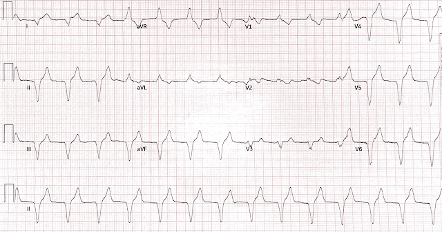
| Sinus bradycardia with PVCs with slight, persistent STE in aVL and I and STD in V4-V6 |
Based on the ongoing ischemic symptoms and persistent STE, I contacted the cardiologist for rescue PCI, which he underwent approximately 3 hours after receiving thrombolytics.
 |
| Coronary angiogram after single drug eluting stent deployment with resulting TIMI III flow |
Troponin I peaked at 14.26 ng/mL 24 hours after arrival to the receiving hospital, consistent with values seen in STEMI. A formal echocardiogram was performed shortly after PCI which demonstrated an EF of 20-25% with akinesis of the anterior wall. Just prior to discharge, a follow up echo was performed which demonstrated improvement in EF to 40-49% and hypokinesis of the anterior wall.
This illustrates the concept of "stunned myocardium." Frequently, the mechanical effects of ischemia persist long after the ischemia is resolved; this may last hours to days to weeks, depending on the duration and severity of ischemia, but ONLY if the myocardium remains viable.
There are times when intervention, or CABG, is high risk and it is only worth performing when the myocardium is viable; such "viability studies" can be done with MRI. In this case, of course that is unnecessary, as PCI was not high risk.
He had an uneventful hospital course and was discharged to home two days after PCI.
This case highlights again how the false dichotomy of the STEMI/NSTEMI paradigm fails our patients. We know that in approximately 25% of NSTEMI cases an OMI will be found at the time of cardiac cath and the short and long term morbidity and mortality of these patients is double that of their STEMI counterparts. [2] In this case, this false decision making tree’s limitations were laid bare: the physician at the outside hospital was forced to choose between STEMI (which it was not) and No STEMI, which would not warrant thrombolytic therapy in the current treatment paradigm. However, this person overcame this bias and did the correct thing for the patient, and undoubtedly spared him a larger MI and all the complications accompanying it.
Teaching points:
STEMI/NSTEMI paradigm is a false dichotomy and should be replaced by the OMI/NOMI paradigm and subjective ECG interpretation
Serial ECGs are useful in the detection of OMI
Bedside ultrasonography may identify regional wall motion abnormalities, which can heighten suspicion for OMI. The wall motion abnormality can be profound, as in this case, or more subtle.
AIVR is a classic finding following reperfusion of an OMI. It is often self limiting, benign, and requires no specific therapy
Thrombolytic therapy is not a contraindication to emergent mechanical reperfusion in the cath lab


Feedback should be brought to the EMS crew regarding AIVR and the (luckily not) possible deleterious treatment with Amio/lido/et al. As lytics are all but gone from most field EMS agencies, many paramedic programs are leaving out the recognition of reperfusion arrhythmias, and the paramedics who remember these arrhythmias are becoming fewer.
ReplyDeleteGood point Christopher. I'll pass your comment on to Alex.
Delete