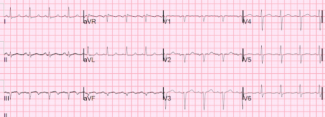
This ECG was presented in a conference.
The patient had presented many times for SOB. On this occasion he was intubated for respiratory failure due to presumed asthma.
I was asked to interpret his ECG in the conference. I had never seen it before.
ECG-1:
This is what I said: "This is diagnostic of an acute inferior MI. There is upsloping ST elevation in III, with reciprocal ST depression in aVL. You do NOT see this in normal variant STE, nor in pericarditis. The only time you see this without ischemia is when there is an abnormal QRS, such as LVH, LBBB, LV aneurysm (old MI with persistent STE) or WPW."
The patient had presented many times for SOB. On this occasion he was intubated for respiratory failure due to presumed asthma.
I was asked to interpret his ECG in the conference. I had never seen it before.
ECG-1:
 |
| What do you think I said? Here is the computer interpretation: (Veritas algorithm) |
This is what I said: "This is diagnostic of an acute inferior MI. There is upsloping ST elevation in III, with reciprocal ST depression in aVL. You do NOT see this in normal variant STE, nor in pericarditis. The only time you see this without ischemia is when there is an abnormal QRS, such as LVH, LBBB, LV aneurysm (old MI with persistent STE) or WPW."
Years later, I ran it through the queen of hearts, and here is her answer:
The Queen of Hearts PM Cardio App is now available in the European Union (CE approved) the App Store and on Google Play.
For Americans, you need to wait for the FDA. But in the meantime:
YOU HAVE THE OPPORTUNITY TO GET EARLY ACCESS TO THE PM Cardio AI BOT!! (THE PM CARDIO OMI AI APP)
If you want this bot to help you make the early diagnosis of OMI and save your patient and his/her myocardium, you can sign up to get an early beta version of the bot here. It is not yet available, but this is your way to get on the list.
Case continued
This OMI was not seen by the providers.
The patient was managed in the ICU and had serial troponins. He had no more ECGs recorded.
Here is the patient's troponin I profile:
Here is the patient's troponin I profile:
 |
These were interpreted as due to demand ischemia, or type II MI.
First was 2.9 ng/mL and subsequentle dropped to 1.5 ng/mL
First was 2.9 ng/mL and subsequentle dropped to 1.5 ng/mL
Such high troponin I is very unusual in type 2 MI.
Here is data from a study we published in 2014 for type II NonSTEMI:
Here is data from a study we published in 2014 for type II NonSTEMI:
Sandoval Y.
Nelson SE. Smith SW. Schulz KM. Murakami M.
Pearce LA. Apple FS. Cardiac Troponin Changes to Distinguish Type
1 and Type 2 Myocardial Infarction and 180-Day Mortality Risk. Acute
Cardiovascular Care 2014;3(4):317-325.
Here is a figure from that study:
| Notice that the 6 hour value (far right) is very low for type 2 MI. |
Cases like this lead me to wonder if troponin should be a vital sign. See this case:
Should Troponin be a Vital Sign? Perhaps, but only if Interpreted Using Pre-test Probability.
After admission to the hospital, the patient was discharged from the hospital without any investigation of his acute MI.
He returned 5 days later for another episode of SOB, which was again diagnosed as asthma (and probably he also had asthma).
ECG-2
I was not involved, but happened to be doing formal reads of ECGs and saw this one in the system:
ECG-3
Here is my interpretation:
| |
|
Management on this 2nd presentation:
The troponin was 0.224 ng/mL and falling, and thought to be due to demand ischemia again [though they were merely coming down after previous (type 1 MI)].
He was discharged and returned again.
At some point he returned with chest pain, and all these findings were put into place.
An angiogram confirmed ACS as the etiology. At this point, all vessels were open and the patient was treated medically (with dual antiplatelet therapy).
Learning Points
1. Again, a computer "Normal ECG" is often abnormal.
2. Troponin I greater than 1.0 ng/mL is seldom a result of demand ischemia (type 2 MI). Beware of diagnosing type 2 MI.
3. Many MI do not have chest pain
4. ANY AMOUNT of STE in lead III, with ANY amount of STD is aVL, in the absence of old MI (inferior LV aneurysm), WPW, LVH, or LBBB is due to acute MI until proven otherwise.
===================================
Comment by KEN GRAUER, MD (3/11/2019):
===================================
Excellent teaching case presented by Dr. Smith regarding how easy it is to overlook an acute STEMI when the history does not suggest this diagnosis, and ECG findings are subtle.
- I focus my comments on the first 2 tracings shown in this case — which for clarity, I have put together in Figure-1.
 |
| Figure-1: The first 2 ECGs shown in this case (See text). |
====================
COMMENT: As per Dr. Smith — ECG #1 was the initial tracing on this patient who presented to the ED already intubated for respiratory failure. The computerized interpretation for this tracing was, “Sinus rhythm; Normal ECG” — and attention of acute care providers was apparently focused on attending to this patient’s pulmonary problems. I would emphasize the following points about this case:
- In my opinion — it is not the fault of the computer that the diagnosis was missed. Instead — it is the fault of the provider who accepts the computer interpretation without independently interpreting the ECG before looking at what the computer said. We have addressed this issue on many occasions in Dr. Smith’s ECG Blog. My views may differ from others — in that as an Attending charged with overreading ECGs for numerous providers — I loved the computerized interpretation once I appreciated what the computer can and cannot do. That’s because the computer saved me LOTS of time (!) by greatly speeding up my interpretation, when I would be confronted with a large stack of ECGs in front of me to interpret. But for anyone who has read less than many thousands of tracings — it is imperative not to even look at the computer interpretation until after YOU have independently interpreted the ECG yourself! Failure to follow this advice will most likely lead to overlooking subtle acute MIs, as occurred in this case. NOTE: I expand on my approach to Optimal Use of Computerized ECG Reports HERE.
- It should be appreciated that the technical quality of ECG #1 is clearly suboptimal. That’s because there is much baseline artifact in each of the limb leads. In my experience, many clinicians fail to acknowledge the presence of obvious artifact — which can make it dramatically more difficult to recognize subtle acute findings. I’ll emphasize that neither Dr. Smith nor myself had any trouble spotting signs of recent MI in ECG #1 despite the artifact — BUT— when artifact impedes interpretation, and ECG findings are subtle (as they are in ECG #1) — it is best to have a low threshold for repeating the ECG as soon as this is feasible. Realizing that you may not be able to eliminate all artifact in an acutely ill patient in marked respiratory distress: i) It is worth a try at a 2nd attempt to get a better quality ECG; and, ii) If an acute cardiac event is in progress — then repeat ECG a few minutes later might now show signs of acute evolution, even if lots of artifact is still present.
Dr. Smith noted in his Learning Points about this Case that, “Many MIs do not have chest pain”. Data on this concept of “Silent” MI was first described in the original Framingham Studies. It is worthwhile keeping in mind important findings from this study:
- More than 1/4 of all MIs are not accompanied by chest pain.
- Of this group (ie, which is comprised of >1/4 of all MIs) — about HALF have NO chest pain at all. The other half ( = >1/8 of all MIs) — have “something else” (ie, some other “non-chest-pain-equivalent” symptom). Among the list of “other non-chest-pain-symptoms” are shortness of breath — GI pain — mental confusion — weakness — myalgias (as in a “flu-like” syndrome) — and simply, “not feeling good”.
- The most common “non-chest-pain-equivalent” symptom is shortness of breath. This is the reason why through the years I always got an ECG on any middle-aged or older adult patient presenting for emergency care with shortness of breath.
Abnormal Findings in ECG #1 — It should be appreciated that no less than 8 of the 12 leads in ECG #1 show abnormalities. So, while the most concerning leads in ECG #1 are leads III and aVL — it is the combination of all of the leads in this tracing with abnormal findings that most heightened my concern. PEARL: Be sure that you always look at all 12-leads on the tracing. Doing so will often “tell a story” by the composite “theme” of abnormal findings.
- Although difficult to see through the artifact — each of the 3 inferior leads (ie, leads II, III, aVF) show ST segment coving (ie, “frowny”-configuration). As already noted — lead III is the most abnormal of the inferior leads, because it already shows T wave inversion. But, straightening with tendency to downward coving of the ST segment in the other 2 inferior leads (as seen in ECG #1) is not normal.
- Although tiny in amplitude, and marred by artifact — lead aVL is suggestive of mirror-image ST depression to the ST coving we see in lead III.
- The other lateral limb lead ( = lead I) shows subtle flattening of the ST segment, with a hint of ST depression. As occurs in this case — reciprocal ST depression in association with acute inferior MI is almost always more marked in lead aVL than in lead I — but in the context of the abnormal appearance of lead aVL in ECG #1 — the ST segment in lead I is not normal.
- Typically, there is slight concave-up (ie, “smiley”-configuration) ST elevation of 1-2 mm in leads V2 and V3 in normal tracings (See Panel B in Figure-2). Instead — the ST segment (before the T wave) is flatter-than-it-usually-is in both leads V2 and V3 of ECG #1, even though there isn’t frank ST segment deviation. In the context of the abnormal limb lead findings described above — the ST segment (especially in lead V2) is not normal, and to me suggests possibly acute posterior involvement.
- Finally — the T wave in lead V6 of ECG #1 is tiny. This is unusual — and in the context of abnormalities in the other 7 leads — the T wave in lead V6 is not normal.
====================
Interpretation of ECG #1: Given the clinical history in this case (ie, No possibility of finding out IF the patient had chest pain — because the patient was already intubated with acute respiratory failure) — I would have interpreted ECG #1 as showing possible infero-postero MI of uncertain age. As an isolated ECG (and, in the absence of a history of new chest pain) — I thought it impossible to “date” this potential MI.
- The point to emphasize is that the term, “of uncertain age” means that IF there is an MI, that it might be an old MI — or, that it might be a recent MI, that could still be evolving. Clinical correlation + serum troponin values and follow-up ECGs will be needed to sort this out.
====================
Interpretation of ECG #2: With recording of the 2nd ECG in this case — it is now readily apparent that this patient is in fact evolving an acute inferior MI because:
- The artifact impeding interpretation of ECG #1 has resolved — and it is now much easier to interpret the abnormal limb lead findings. There are new large Q waves in leads III and aVF of ECG #2 — with a straightened ST segment takeoff of the elevated ST segments in these leads + T wave inversion now also in lead aVF as well as lead III.
- It is much easier in ECG #2 to appreciate the mirror-image opposite reciprocal ST-T wave changes in lead aVL, with respect to the ST elevation we see in lead III.
- It is also much easier in ECG #2 to appreciate some reciprocal ST depression in lead I.
- Finally — there has been further evolution of the subtle ST-T wave abnormalities in leads V2, V5 and V6 — which now all show definite ST-T wave flattening in ECG #2.
 |
| Figure-2: Descriptive analysis terms regarding ST elevation (See text). |
====================
ADDENDUM: Descriptive analysis of ST-T wave changes can be confusing. The terms, “concave up” and “convex down” are often used to describe the shape of elevated ST segments.
- I was taught that the shape of the ST segment in Panel B of Figure-2 reflects ST elevation with an upward concavity — as compared to the shape of the ST elevation in Panel A, which is convex down.
- That said, whether the ST segment is described as concave up or convex down really depends on whether the viewer is looking from above or below the ST segment. Use of this terminology can be confusing ...
- For clarity when communicating among colleagues — I favor use of the descriptive terms, “smiley” configuration (which would be concave up, by the terminology I was taught) — and “frowny” configuration (which corresponds to convex down by the terminology I was taught).
- I also like the descriptive term, “coved” — to reflect a “frowny”-shaped ST segment.
- Clinically — the importance of ST segment SHAPE cannot be overstated! In general — smiley-shape ST elevation is more likely to be benign (as is commonly seen with repolarization variants) — whereas ST segment coving is more likely to reflect an acute process. NOTE: Many exceptions to this generality exist! Space constraints here don’t permit full discussion of these exceptions. My point is simply that ST segments in each of the inferior leads of ECG #1 (shown in Panel C of Figure-2) suggest a tendency to ST segment coving, which is not a normal ST segment shape in the inferior leads. This is best seen by the imaginative “frowny” configuration, which I have superimposed on the magnified lead II ST segment.


This is an example of subtlety at its finest! And great discussions.
ReplyDeleteChronic pulmonary patients can be very difficult to diagnose because 1) as in this case, cardiac compromise may present as shortness of breath instead of chest pain, 2) when they do have chest pains, they are often due to non-cardiac factors and 3) their mean QRS axis is almost always very posterior making it more perpendicular to the frontal plane. This results in small (more difficult to interpret) deflections in the limb leads and larger deflections in the precordial leads.
Whenever you see deflections that appear unexplainable on the ECG of a chronic lung patient, don't forget that the mean QRS axis exists in THREE - not TWO - dimensions and that every axis has TWO perpendiculars - not just ONE!
Regarding the convex up/concave down dilemma, I just think of everything as "up": "concave up" or "convex up." I never use the word "down."
Nice points — Thanks Jerry! — :)
Delete