Wednesday, February 28, 2018

ST-Elevation in aVR with diffuse ST-Depression: An ECG pattern that you must know and understand!

This case comes from Sam Ghali  (@EM_RESUS). 

A 60-year-old man calls 911 after experiencing sudden onset chest pain, palpitations, and shortness of breath. Here are his vital signs:

HR: 130-160, BP: 140/75, RR:22, Temp: 98.5 F, SaO2: 98%

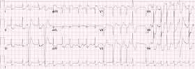
This is his 12-Lead ECG:

He is in atrial fibrillation with a rapid ventricular response at a rate of around 140 bpm. There are several abberantly conducted beats. There is ST-Elevation in aVR of several millimeters and diffuse ST-Depression with the maximal depression vector towards Lead II in the limb leads and towards V5 in the precordial leads.


ECG reading is all about pattern recognition. And this particular pattern of ST-Elevation in aVR with diffuse ST Depression is a very important ECG pattern that you must be able to recognize. But what's probably more important than being able to recognize the pattern, is understanding what it represents. There appears to be a common misconception that the ST-Elevation in aVR in this case possibly represents "STEMI", or acute transmural (full-thickness) ischemia. If this were the case the patient would most likely be dead or at the very least in profound cardiogenic shock. The key to understanding what this pattern represents lies in understanding that the ST-Elevation in aVR is reciprocal to the diffuse ST-Depression - and that this diffuse ST-Depression represents global subendocardial ischemia!


So the real question that you must answer is: 
What is causing the global subendocardial ischemia?


It is critical to realize that more often than not the cause is global myocardial strain from a Non-ACS etiology! (profound sepsis, tachycardia, anemia, hypoxemia, etc). It is also very important to understand that in these Non-ACS settings, you can see this pattern with or without underlying coronary artery disease.

But of course it could be ACS. And if it is, then you are dealing with Left Main, Proximal LAD, or even multi-vessel plaque instability. But keep in mind that even if it is ACS you are still dealing subendocardial and not transmural ischemia.


Here is a subcostal view of the bedside Echo obtained from our patient in the ED:
There is good global function


So what is causing the diffuse subendocardial ischemia in our patient? 

 When the heart rate is significantly elevated as in this case, it is reasonable to suspect that the ischemia is likely tachycardia-induced, or "demand ischemia." So given the normal EF noted on Echo, (and by the way I would strongly recommend assessing the EF of any patient before deciding to give any negative inotropic medications) the decision was made to administer a Diltiazem bolus and infusion and to reassess after rate control was established. Rate controlled was gained as the patient's heart rate came down very nicely into the 80's. He felt much better and his symptoms were all but completely relieved.


Smith comment: it is also critical to assess volume before giving negative inotropes and negative chronotropes.  This tachycardia could be a response to poor LV filling.  Indeed the neither LV nor RV appear to be filling very well.  If the atrial fib with RVR is resulting in a rate so fast that the rate is the cause of poor LV filling, then there should be some increased filling pressures, possible pulmonary edema, and evidence of fluid overload.   Assessment of IVC filling would be helpful, and, if it is collapsed, then administration of fluids first (or blood if this is a GI bleed) is indicated.  If this does not result in a slower heart rate, then an AV nodal blocker is indicated, such as Diltiazem.  

Furthermore, since this patient has no history of atrial fib, and it is a critical situation, electrical cardioversion is both safer and more effective than an AV nodal blocker such as Diltiazem.

See these 2 posts

Atrial fibrillation with RVR: use POCUS to assess volume; then sinus vs. SVT: use of Lewis leads


Here is the repeat ECG obtained 25 minutes after the first one:
The rhythm is still A-Fib. The heart rate has come down more than 65 points. But despite the dramatic decrease in heart rate, the pattern of global subendocardial ischemia persists! (ST-Elevation in aVR with diffuse ST-depression that has a maximum depression vector towards leads II and V5)


If this repeat ECG had shown resolution of the global subendocardial ischemia pattern, it would be reasonable to conclude that it was likely the result of a-fib with an uncontrolled ventricular response. But because this pattern persisted after rate control and in the absence of any other evidence of clinical causes, one must assume that the etiology of the pattern is indeed ACS - meaning there has been acute plaque instability in either the left main coronary artery, Proximal LAD, or multi-vessel involvement. 

The patient was started on heparin in addition to the aspirin he received en route and the Cardiology team was consulted. (Of note, it is important not to start these patients on dual anti-platelet therapy as there is a high likelihood that they will require CABG.) The decision was made to proceed urgently to the cath lab for angiography. 


Cardiac Cath Results:

Left Main: There is a 90-95% stenosis of the distal left main including the ostium of LAD and Left Circumflex arteries.  
LAD: There is a focal 80% stenosis just after the takeoff of the first diagonal branch.

Circumflex: Severe disease at its ostium and moderate disease in the remainder of the proximal segment.

RCA: 100% chronic total occlusion at its proximal segment.


In discussion with the interventional cardiologist who performed the cath there was thought to be evidence of likely a component of acute thrombus at the 90-95% left main stenosis, suggesting Left Main ACS!


Case Resolution:

The patient was referred for CABG and ended up doing quite well.


Take Home Points:

1. The key to ECG reading is pattern recognition. The pattern of ST-Elevation of at least 1mm in lead aVR + diffuse ST-Depression with a maximal depression vector towards leads II & V5 is a pattern you must know. It represents global subendocardial ischemia.

2. When you see this pattern you should divide the differential for the diffuse subendocardial ischemia into two main categories: ACS vs Non-ACS. Do not automatically assume that it is ACS. I have seen this mistake made many times as ACS becomes the focus, at the expense of appropriate resuscitation addressing the underlying cause. It is very important to keep in mind that the etiology is far more likely to be Non-ACS than ACS!

See this case: 

Diffuse Subendocardial Ischemia on the ECG. Left main? 3-vessel disease? No!


3. The key to determining the etiology is through history, physical exam, clinical picture, laboratory data, Echo, and vigilant monitoring and frequent reassessment.  If you have identified and addressed potentially reversible causes of the ischemia, and the ECG pattern persists then you are dealing with ACS until proven otherwise.

4. Refrain from using dual-antiplatelet therapy in these patients as there is a high likelihood they will require CABG.

5. Remember that if this ECG pattern does represent ACS, the ST-Elevation in aVR is not the result of direct injury (or transmural ischemia) and that the ST-Elevation in aVR is reciprocal to the diffuse ST-Depression. Therefore these ACS cases do not represent "STEMI".  However, while there is not great data to guide the timing of cath for these patients, I would advocate going to the cath lab with a much stronger sense of urgency than for other "NSTEMIs".  The reasoning is that ACS is a very dynamic process and without the advantage of optimal medical therapy (a second platelet inhibitor should be withheld) there is a higher chance of the culprit vessel suddenly occluding and evolving to transmural ischemia. If this happens in the Proximal LAD, Left Main, or in the setting of Multi-vessel involvement the myocardial territory in jeopardy is so large that there is a good chance the patient will arrest and die before any reperfusion can be established!

6.  Smith comment: With diffuse subendocardial ischemia, you may not see any wall motion abnormality.  Global function can even be normal, although it may be globally depressed as well.  A normal bedside echo does not help in: 1) differentiating the cause of the STE in aVR 2) ruling out ACS.


Here is the section on aVR written by Smith in: Miranda et al. "New Insights into the Use of the 12-lead ECG in Acute MI in the ED" (Canadian J Cardiol 34(2):132-145; Feb 2018)


Lead aVR in ACS 62

Many experts consider the ECG pattern of STE in aVR, with diffuse STD elsewhere (referred to herein as the aVR STE pattern), to be representative of LM ATO.7 The 2013 ACC/AHA STEMI guidelines consider this a STEMI equivalent,in which thrombolytic therapy is not contraindicated (evidence level B, no specific class of recommendation).  18 However, these conclusions are on the basis of studies in which LM lesions were not true subtotal or complete occlusion (ie, TIMI 0/1 flow).62,63 The interventional community defines occlusive LM disease as >50% according to fractional flow reserve, or 75% stenosis,64 but urgent or emergent intervention on lesions not meeting these thresholds is only imperative if it is a thrombotic lesion and the patient has refractory ischemic symptoms (ie, not resolved by nitrates, antiplatelet, and antithrombotic therapies; see 3 examples in Supplemental Fig. S7).

Although nearly half of patients with 1 mm STE in aVR due to ACS will require coronary artery bypass surgery for revascularization,62 the infarct artery is often not the LM, but rather the LAD or severe 3-vessel disease. More importantly, such ECG findings are frequently due to nonocclusive etiologies (eg, baseline LVH, demand ischemia secondary to respiratory failure, aortic stenosis, hemorrhagic shock). Knotts et al. reported that only 23% of patients with the aVR STE pattern had any LM disease (fewer if defined as 50% stenosis). Only 28% of patients had ACS of any vessel, and, of those patients, the LM was the culprit in just 49% (14% of all cases).57 It was a baseline finding in 62% of patients, usually due to LVH.

Thus, a number of expert reviews emphasize the low specificity of the aVR STE pattern, preferring to label it as circumferential subendocardial ischemia; in this syndrome, STE in aVR is reciprocal STE, reciprocal to an STD vector toward leads II and V5.10,12,62 

The aVR STE pattern is also not sensitive for LM ATO. However, anterior STEMI with combined new right bundle branch block and left anterior fascicular block is highly suggestive of LM ATO (see example 12-lead ECG in Supplemental Fig. S8).65,66

It should be re-emphasized that true LM ATO (ie, TIMI flow 0) is rare in the ED, because most either die before arrival or are recognized clinically because of cardiogenic shock. Thus, reported specificities of STE in aVR for LM ATO result in very low positive predictive values. Of those who do get to the ED, many present with clear STE.62,65,66

The ACC/AHA states that thrombolytics are not contraindicated for diffuse STD associated with  STE in aVR.  Because of the poor specificity of this pattern for LM ATO, we suggest that thrombolytics should only be considered for those with profound STD that is clearly due to ACS, is refractory to all other medical management, and only when PCI is completely unavailable.

Lead aVR in STEMI
 Some patients whose ECGs already meet conventional STEMI criteria might also have STE in lead aVR. This finding does not alter the need to pursue emergent reperfusion, although it might suggest a poorer prognosis.62,67  In a patient with otherwise diagnostic STE, additional STE in aVR does not represent LM ATO and is not helpful in diagnosing the infarct-related artery or the site of occlusion.68  Less than 3% of anterior STEMI has LM ATO, and most are recognized clinically because of cardiogenic shock.69,70


References


62. Smith SW. Updates on the electrocardiogram in acute coronary syndromes.  Curr Emerg Hosp Med Rep 2013;1:43-52.

63. Jong GP, Ma T, Chou P, et al. Reciprocal changes in 12-lead electrocardiography
can predict left main coronary artery lesion in patients with acute myocardial infarction. Int Heart J 2006;47:13-20.

64. Stone GW, Sabik JF, Serruys PW, et al. Everolimus-eluting stents or bypass surgery for left main coronary artery disease. N Engl J Med 2016;375:2223-35.

65. Fiol M, Carrillo A, Rodriguez A, et al. Electrocardiographic changes of ST-elevation myocardial infarction in patients with complete occlusion of the left main trunk without collateral circulation: differential diagnosis and clinical considerations. J Electrocardiol 2012;45:487-90.

66. Widimsky P, Rohac F, Stasek J, et al. Primary angioplasty in acute myocardial infarction with right bundle branch block: should new onset right bundle branch block be added to future guidelines as an indication for reperfusion therapy? Eur Heart J 2012;33:86-95.

67. Kukla P, Bryniarski L, Dudek D, Krolikowski T, Kawecka Jaszcz K. Prognostic significance of ST segment changes in lead aVR in patients with acute inferior myocardial infarction with ST segment elevation. Kardiol Pol 2012;70:111-8.

68. Kosuge M, Ebina T, Hibi K, et al. An early and simple predictor of severe left main and/or three-vessel disease in patients with non-ST-segment elevation acute coronary syndrome. Am J Cardiol 2011;107:495-500.

69. Zoghbi GJ, Misra VK, Brott BC, et al. ST elevation myocardial infarction due to left main culprit lesions: percutaneous coronary intervention outcomes. J Am Coll Cardiol 2010;55:A183.E1712.

70. Kurisu S, Inoue I, Kawagoe T, et al. Electrocardiographic features in patients with acute myocardial infarction associated with left main coronary artery occlusion. Heart 2004;90:1059-60.


Saturday, February 24, 2018

Right Bundle Branch Block and ST Depression in V1-V3. Is that normal? And a complication.

A late middle-aged male with h/o 3 vessel bypass (CABG), type 2 diabetes, peripheral vascular disease, DVT, Chronic Kideny Disease, and chronic combined systolic and diastolic congestive heart failure presented with chest pain which started approximately 2 hours prior to arrival.

Here was the initial ED ECG:
Sinus Rhythm
There is an rSR' in V1, with wide S-waves in lateral leads (right bundle branch block, RBBB).
Normally, RBBB has a bit of ST depression in V1-V3 that is discordant (in the opposite direction of) the R'-wave.
So that bit of ST Depression in V1 is normal.
What about V2 and V3?


Notice there is no R'-wave in V2 and V3!!  This happens in some RBBB when there is very early transition.  Normally, in RBBB, there is indeed an R'-wave in V1-V3.  Here it is only in V1, and the wide S-wave which is normally seen in V4-V6 starts all the way rightward at V2.  Therefore, one should not expect any ST depression in V2.


But this was made easier because there was a previous ECG available:
There is no ST depression in V2 and V3 on this old ECG
Also, there was no STD in V1 either


No action was taken, but a second ECG (below) was recorded 80 minutes after the first. In the meantime, a nitro drip had been started and aspirin and heparin given.

This one was texted to me with the words: "Ongoing improving pain with trop 0.13 (elevated).  Creatinine 2.2."

T=80 minutes:
I could only see it on my phone, and I had no other clinical info.  I responded:
"An unusual RBBB, but there is quite a bit of ST depression in V2 and V3.  Must assume it is ischemic.  Must go to cath lab unless it resolves"

When I had a chance to view these on a full screen, it became clear that this is all but diagnostic of posterior STEMI.   Not all ST depression in V2 and V3 is posterior STEMI, but it is posterior STEMI until proven otherwise.

Note: posterior leads do not help here: no matter what the cause of the ST depression in V2 and V3, the posterior leads will reflect the opposite! This is an electrical necessity!  They will not be able to determine the cause of the ST shift.

It is well known that the most likely cause is posterior STEMI. 

When should you record posterior leads?  If you suspect ischemia and it is not showing on the 12-lead, on occasion it will manifest in posterior leads only.

The patient's pain completely resolved on a nitro drip.

He was admitted.

There was a 3rd ECG at 240 minutes:
ST depression is mostly resolved, but the ECG has not returned to baseline.

A 4th ECG was recorded at 360 minutes:
No difference from 240 minutes


The patient had positive troponins and was scheduled for angiography to begin about 16 hours after arrival in the ED.

Complication: While waiting in the cath lab prep room, he had VT and V Fib arrest.

Fortunately, he was easily defibrillated.

Here is the 12-lead recorded immediately after resuscitation:
There is sinus bradycardia with a junctional escape.
RBBB persists
Now there is profound ST depression in V1 and V2

Angiogram:  it is complex because of previous CABG, but the bottom line was that there was an acute 99% thrombotic occlusion of the saphenous vein graft to the obtuse marginal (to the posterior wall).

EchoDyskinesis of the basal to mid inferior and inferolateral segments.  (Basal inferior segment is equivalent to the posterior wall).  EF 45% (not significantly changed from prior).

Peak troponin I: only 2.94 ng/mL.

The patient did well.  Here are subsequent ECGs:

Immediate post cath ECG:
There is trigeminy: complexes 2, 5, 8, and 11 are all PVCs (each is in the middle of the 3 complexes in each of (I, II, III / aVR, aVL, aVF / V1-V3/ V4-V6).  
ST depression is mostly resolved.


ECG at 36 hours:
Some persistent ST depression.  

ECG at 60 hours:
All ST depression has resolved.


Summary

The patient was fortunate to have a STEMI with delayed treatment that did not result in a lot of myocardial loss.

Final formal diagnosis: NonSTEMI

You can see what a misnomer this is.  It is a STEMI, but with failure to record ST elevation because the 12-lead does not record over the posterior wall.

This is therefore another example of the False STEMI-NonSTEMI Dichotomy
See my lecture on this topic: 
Lecture at the 2015 SMACC Chicago conference:"The False STEMI-NonSTEMI Dichotomy".


Learning Points
1. Ischemic ST depression in V2 and V3 due to ACS is posterior STEMI until proven otherwise.
2. It should be called STEMI
3. RBBB has up to 1mm of normal ST depression in V1-V3, but ONLY when there is an R'-wave!!  That ST depression is rarely more than 1 mm (and then only when the R'-wave is very large, such as in right ventricular hypertrophy)
4. If you treat a patient with a "NonSTEMI" medically, with delayed cath lab, you MUST monitor extremely closely.  Arrest can happen at any time.


Thursday, February 22, 2018

Shoulder pain after lifting a heavy box

Written by Pendell Meyers, edits by Steve Smith


This will be too easy for most long-time readers, but if you are at that level, sit back and enjoy noticing how few milliseconds it takes to recognize this thanks to so many prior examples on this blog!

I was sent this ECG from EMS with only the information that it belonged to a middle aged male with left shoulder pain.
What do you think?


There are hyperacute T-waves in leads V1-V6, as well as in leads II, III, aVF. The J-points are all at baseline with the exception of leads V2-V3 which show a small amount of STD (which makes de Winter morphology in the presence of hyperacute T-waves).

How can you explain that the most obvious findings are in the anterior leads, yet the inferior leads are also hyperacute??

The occluded vessel must supply the anterior wall and also the apex and/or inferior wall. The most common variant that satisfies this is a type III "wraparound" LAD. This is a large and long LAD that wraps around the apex of the heart, supplying the apex and sometimes even parts of the inferior wall.

By ECG, this acute coronary occlusion is predicted to be of very short time duration, with very high acuity and very high viability. As shown in our reference diagram below, hyperacute T-waves generally exist only within a few hours of persistent acute coronary occlusion, or immediately after reperfusion ("on the way up, and on the way down," as Dr. Smith says).


We activated the cath lab based on this EMS ECG, because it is obviously diagnostic of acute coronary occlusion involving the anterior, lateral and inferior walls. When I make this decision prospectively on this particular highly diagnostic ECG, I estimate that the likelihood of acute coronary occlusion as the etiology of these ECG findings is approximately 99%, with the remaining 1% being the occasional takotsubo cardiomyopathy with indistinguishable ECG findings, and which can only be differentiated by angiogram.


The patient arrived in the resuscitation bay at the same time as the cardiologist.

He was a middle aged man with history only of HTN who called EMS for "soreness" of the left shoulder while working in his garage. He stated he lifted a box weighing approximately 75 lbs, then set it back down, then noticed severe pain in his left shoulder described as "soreness" and "pressure." He stopped working, but the pain persisted. He waited 2-3 hours at home before calling EMS thinking the pain might simply go away.

Here is his initial ED ECG:
Essentially the same findings, hyperacute T-waves without dramatic ST segment changes.


The cardiologist was somehow not impressed by these findings. He also thought that the pain was musculoskeletal because it started around the time of lifting a heavy box. Yet on exam the patient had full range of motion without any change in his constant severe shoulder pain.

I advised the cardiologist that this patient must be taken immediately for cath and intervention. He stated that this ECG does not meet STEMI criteria. I said that the patient has an acute coronary occlusion based on the hyperacute T-waves, the same pathology as an obvious STEMI. The only difference being that there is even more viable myocardium to save than a classic obvious STEMI because there is not yet STE.

Note: The reason there is even more myocardium to save than classic STEMI is because acutely ischemic myocytes first "register" in the T-wave and create increased area under the T-wave, then as they start undergoing the process of death they register in the ST segment, and finally when they are stunned or dead they cannot conduct the action potential and register in the Q-wave. As far as I know this is not proven on a cellular level but is well supported by my experience and hundreds of cases on this blog.

He asked me where I thought the lesion was based on the ECG, and I said "mid LAD or higher, and the LAD will be a type III wraparound."

I stood by the monitor, getting repeat ECGs every 5 minutes for the next 20 minutes while trying to convince the cardiologist, expecting the repeat ECGs to show evolution to frankly obvious STE. But the ECGs did not change - hyperacute T-waves were present non-stop for approximately 45 minutes (from EMS ECG to my last ED ECG). The patient stated that his pain had been exactly the same for 3-4 hours, with no episodes of decreasing and then returning pain.

In my experience (and Dr. Smith agrees), it is unusual for hyperacute T-waves to last this long without progression or evolution. Most cases we have on this blog show evolution to obvious ST elevation, or you see the predictable progression of reperfusion and reocclusion with hyperacute T-waves in both directions. It is possible that there was reperfusion and then reocclusion between the EMS ECG and the ED ECG, although this is less likely because the patient denied temporary improvement in symptoms. Interestingly, Dr. Smith notes that de Winter himself stated that his characteristic morphology was stable for several hours, although Dr. Smith's opinion is that de Winter's data did not actually support that assertion.

It is possible that he had some very small source of collateral flow which was just barely enough to prevent progression, keeping him on the upper end of the de Winter pathology spectrum. It is also possible that the patient had recurrent brief episodes of reperfusion and reocclusion which did not have enough time to show the progression of ECG findings before reversing.


My fellow resident performed a bedside US showing a very dense anterior and apical wall motion abnormality, further confirming the diagnosis.


Thankfully, the cardiologist took the patient to the cath lab at this point.

Here's what they found!

100% mid-LAD occlusion.

With red arrows at the site of occlusion.


Mid-intervention, you can see the occlusion has been opened and there is a very large territory of myocardium supplied by the previously occluded LAD.


After intervention with good flow. This LAD is large and long such that it cannot be captured in one frame, and is seen extending down further in other images (not shown), wrapping around the apex of the heart. This confirms wraparound LAD.


Here is his post-cath ECG approximately 2 hours after the above ED ECG (no ECGs available between my ED ECGs and this one):


What do you think?


This ECG shows progression of acute myocardial infarction to almost complete transmural anterior wall loss. There are new deep wide pathologic Q-waves in V1-V5, with persistent STE and some loss of T-wave hyperacuity. Hyperacute T-waves in the inferior leads are now much less hyperacute than seen in the prior ECG.


But we're done, right? The angiogram confirms successful reperfusion, right?

WRONG.

The ECG is much better at confirming or denying reperfusion than the angiogram. The reason is that epicardial large vessel flow does not necessarily ensure that the actual myocytes downstream are receiving blood flow. The epicardial vessel obviously branches into innumerable smaller branches to provide capillary circulation to the cells. The ECG measures the viability of the cells rather than the flow through the epicardial coronary vessels. Expert ECG interpretation is better than angiogram, better than troponin, even better than patient reported symptoms for determining the state of occlusion and reperfusion!

"No reflow" phenomenon is described when a patient seemingly has successful reperfusion on angiogram but persistently progressing myocardial infarction. This is usually evidenced by ECG changes that progress along the occlusion progression below, and looks similar to the progression seen in patients who receive no intervention at all. This is thought to be due to downstream showering thrombi and platelet aggregates into the microcirculation or other unknown pathophysiology.

So knowing that this patient had relatively good door to balloon time, good angiographic result, and hyperacute T-waves just prior to emergent cath, what do you expect to see for his peak troponin over the next 24 hours?


Answer: Very very high, because despite the angiographic result the ECG shows complete infarction.


Indeed, his peak troponin T was 8.89 ng/mL (very highly elevated). Echo showed EF 35% with dense anterior and apical wall motion abnormality.

Here is the next day ECG:
T-waves further deflating as expected in the course of subacute total LAD occlusion infarction. Remember, when this QRS morphology is present the T-wave becomes almost the only reliable indicator of progression/reocclusion/reperfusion, and is in fact the main consideration used in decision aids to differentiate acute LAD occlusion versus persistent STE (LV aneurysm morphology).


These ECGs are unfortuantely an excellent example of the progression to "LV aneurysm morphology" (see below for reference). The ECG will likely remain similar to the above indefinitely. It is critical to recognize this morphology as this patient is at highest possible risk for the classic complications of transmural infarction including anatomic LV aneurysm, mural thrombus with subsequent stroke, free wall rupture, VSD, Dressler's syndrome, etc. He is also at risk of another complication that gets less discussion: misdiagnosis of his new baseline ECG! He will likely have persistent STE which will be alarming to his future providers, yet the T-waves will likely be the most reliable electrocardiographic feature to tell us whether he is experiencing further acute coronary occlusion affecting his anterior wall. He is at risk of mismanagement in both directions. Should he unfortunately suffer a pulmonary embolism or pericarditis or simply GERD or chest wall pain, for example, he may present with chest pain and this ECG, prompting premature diagnostic closure and immediate catheterization rather than further workup for other causes. Should he suffer another acute coronary occlusion, diagnosis may be missed or delayed because his ST segments are not appreciated as different from his baseline ECG which will now have STE forever.


The patient recovered and did well. Long term outcome unknown.


Learning points: 

1) You MUST be able to recognize hyperacute T-waves such as these. They are common early in the course of acute coronary occlusion.

2) You must learn and advocate for your patients, as these ECG findings are not widely taught or known since our current guideline-promoted strategy focuses only on the misguided STEMI criteria. 

3) Hyperacute T-waves and/or de Winter morphology may be present for hours without obvious evolution, but in most cases you can find evidence of progression with serial ECGs. They may even represent complete occlusion with ongoing necrosis (infarction)!

4) Type III Wraparound LAD is a common anatomic variant which produces anterior and inferior findings on ECG.

5) The ECG is the most accurate measure of occlusion and reperfusion, even better than angiogram, laboratory values, or patient symptoms.

6) You must become familiar with the ECG findings of complete full thickness infarction, as well as the clinical syndromes and complications that ensue.


Monday, February 19, 2018

Altered Mental Status, Bradycardia

911 was called for an elderly woman who fell and was confused.  Medics found her unresponsive, with "convulsive" movements.  They could not find a pulse.  They performed CPR, gave epinephrine, and intubated the patient and regained a pulse, at which time she became responsive.

On arrival, heart rate was 87 and she was hypotensive at 52/21, with a palpable pulse and cardiac function present on echo.  She was intubated (by medics), but awake and alert and nodding to questions, shaking her head "no" to chest pain, headache, or SOB.  Repeat pulse was slow and irregular.  She was non-focal and followed commands.

Her heart rate dropped back down and an ECG was recorded:
Thoughts??


First, there are very sublte regular P-waves at a rate just over 100, seen best in lead II across the bottom.  The ventricular response is irregular and dissociated, and narrow, so there is complete AV block with variable junctional escape.

And one other finding.   At conference, one of our smart faculty, Dr. Richard Gray, immediately made the diagnosis without any other clinical information, though it is very subtle.  What is it?

This magnification of lead III may help, though it is still very subtle:
What is that bump at the end of the QRS?


The diagnosis was not suspected based on either clinical or ECG grounds.  The patient was given atropine 0.5 mg x 3 with no response.

Labs were:
VBG: 7.21/59/39/23
Lactate: 4.8
Hgb: 13.7
Na: 138
Cl: 109
K: hemolyzed at 8.1
CO2: 23
Cr: 2.62

After ketamine sedation, transcutaneous pacing was begun at a rate of 70, with capture at 48 milliamps.

It was found that the patient was on metoprolol and diltiazem, so Calcium gluconate 3 grams was given for both possible hyperkalemia and calcium channel blocker (CCB) toxicity.  Glucagon 1 mg was given for possible BB toxicity.

High dose insulin (HDI) was started at 1unit/kg for probably CCB and BB toxicity.

At 60 minutes, transcutaneous pacing was stopped and another ECG was recorded:
The finding is still very subtle.


It is very small Osborn waves.  In the second ECG, it is most apparent in lead V6, though still extremely subtle.

This is a diagnosis that should be made clinically.  One would think that you don't need an ECG to diagnose hypothermia, but sometimes the temperature is not easy to get.

In this case, they had trouble obtaining a rectal temperature.  This alone is a clue that it may be very low.  When they finally did get a temperature by urinary catheter probe, it was 29 degrees C, or 84 degrees F.

In this case, the ECG had atrial fibrillation with a very slow ventricular response.  One should not only suspect hypothermia, but also hyperkalemia and drug toxicity.  All three contributed here.  Transcutaneous pacing and treatment for all 3 disorders was undertaken with success.

Ultimately, the patient was found to be uroseptic.

Tiny Osborn waves.

If you don't believe the finding in lead III, look at this series of ECGs that start at 23 degrees and end at 29 degrees.  Look at lead III on the 4th (last, at 29 degrees) ECG.

Massive Osborn Waves of Severe Hypothermia (23.6 C), with Cardiac Echo


Learning Point:

1. Always obtain an accurate temperature in critically ill patients.  Rectal temp may not record accurately.  If the temp does not record, consider that it is too low.  Or get an EKG (just kidding).

2.  Use transcutaneous pacing when bradycardia results in cardiac output that is insufficient (shock, in this case manifested by ).

Everything you need to know about transcutaneous pacing:


Emergency Transvenous Cardiac Pacing


Thursday, February 15, 2018

A female in her 60s who was lucky to get expert ECG interpretation

Submitted and written by Alex Bracey, with edits by Pendell Meyers and Steve Smith:

I was walking through the critical care section of the ED when I overheard a discussion about the following ECG. I had no history on the case and no prior ECG for comparison.

What do you think?


Here are inferior leads, and aVL, magnified:
A closer inspection of the inferior leads and aVL
Sinus bradycardia. 
The T-wave in lead III is slightly tall and broad (increased area under the curve) compared to its QRS complex. In isolation, this probably could not be called a hyperacute T-wave, but you may suspect it.   
There is T-wave inversion (TWI) in aVL.  

T-wave inversion in aVL: when is it abnormal?

There is no LVH or LBBB on which to blame the TWI (i.e., the QRS is normal). While T-wave inversion in aVL may be normal in the presence of a normal QRS, this is only true when the T-QRS angle is small. That is to say, when the T-axis and QRS axis are similar. In other words, if the QRS is negative, the T-wave may be negative. However, here QRS axis is about 35 degrees and the T-axis is about 85 degrees. Thus the T-QRS angle is 25 - 85 = (-60) degrees, which is abnormal. Any absolote value greater than 45 degrees is suspicious for T-wave inversion (however, this is very complex; see table posted at the bottom of this post.)

Now that we know the T-wave inversion in lead aVL following a normal QRS complex is abnormal, it helps to confirm that the T-wave in lead III is indeed hyperacute. The flattened T-wave in V2 suggests likely posterior involvement.

The cath lab was not activated based on this initial ECG. The patient was a female in her 60s with history of HTN and smoking who presented for chest pressure x1 hour. The initial ECG was taken at 0839. Based on the initial ECG and presenting complaint, the attending involved in the case opted to keep the patient in our critical care unit for close monitoring and serial ECGs.


She went on to describe her chest pain as a "buffalo sitting on my chest" and a "weird" sensation in her jaw for 1 hour prior to arrival, associated with lightheadedness and diaphoresis.


The patient was given fentanyl initially for chest pain with minimal effect and then vomited which was followed by zofran and famotidine. Initial troponin T from 0840 was less than 0.010 ng/mL (undetectable).


The following ECG was obtained at 0910:


Repeat ECG recorded 30 minutes after initial ECG. The patient still had chest pain.
Magnified:
Again, a close up of the interior leads and aVL


Sinus rhythm with borderline 1st degree AV block. There has been interval marked increase in the area under the ST-segment and T-wave in leads II, III, and aVF, with concomitant increase in the area above the inverted T-wave in aVL, all confirming that that these truly represent inferior hyperacute T-waves. The STE in the inferior leads is larger. There is now TWI and small STD in lead V2, highly suspicious for posterior MI. 


This ECG is diagnostic of an acute coronary occlusion of an artery supplying the inferior and posterior walls.


The cath lab was activated following this ECG and the cardiology fellow came to the bedside. The patient was given aspirin and ticagrelor and was scheduled for urgent cath.


In the cardiac cath holding area, a repeat troponin T was 0.01 ng/mL (positive). The following ECG as recorded at 1206:


Pre-cath right sided ECG. V1 and V2 are unchanged from the normal 12-lead sytem; V3-V6 are actually V3R-V6R.
The T-waves are even more hyperacute. Right sided leads (V3-V6 on this ECG correspond to V3R-V6R) have STE and hyperacute T-waves indicative of RV infarction.

Progression of Inferior leads and aVL

In this magnified arrangement you can see progression of subtle changes including the progressively increasing area underneath the hyperacute T-waves. Even at ~4 hours into her acute coronary occlusion there is barely any ST Elevation. 

Also notice that the ST segments in this example are concave, which is often erroneously mythologized as a non-ischemic pattern.  


Progression of V2 showing posterior involvement.


The patient was then taken to the cath lab an found to have a proximal RCA 100% thrombotic occlusion which was successfully stented.


100% occluded RCA with TIMI 0 flow

Post drug-eluting stent placement with TIMI 3 flow


While in the cath lab, she transiently developed complete heart block and became hypotensive requiring transvenous pacemaker placement and transient pressors. A right heart cath revealed increased right heart pressures and a similarly timed echo revealed mild right heart failure.

Peak troponin T was 3.00 ng/mL (highly elevated).

Post-cath ECG with resolution of acute changes.


The transvenous pacemaker was removed the following day and pressors were not required again. She was discharged to home 2 days later without further complications.

Learning Points:

1) As we have previously demonstrated, aVL was once again the key initial clue to diagnosing subtle RCA occlusion.

2) Contemporary troponins only start to rise 4-6 hours after the onset of acute coronary occlusion. Relying on troponin elevation to diagnose acute coronary occlusion after at least 4 hours of infarction when the ECG can identify it immediately is poor choice.

3) STEMI criteria failed to identify this acute coronary occlusion, like many others. Only expert ECG interpretation combined with strong clinical suspicion were able to identify this case. Remember that some acute coronary occlusions will present with totally normal serial ECGs, and some patients with extremely concerning symptoms warrant emergent cath lab activation even without ECG findings.


QRST Angle

New abstract on QRST angle:


Strebel I et al.  Diagnostic and prognostic value of the QRS-T-angle, an ECG marker quantifying heterogeneity of depolarization and repolarization, in patients with suspected non-ST-elevation myocardial infarction.  J Electrocardiology January–February, 2018; Volume 51, Issue 1, Pages e5–e6.

Background: The value of the 12-lead ECG in the diagnosis of non-ST-elevation myocardial infarction (NSTEMI) is limited due to insufficient sensitivity and specificity of standard markers of ischemia and because ECG confounders may prevent their application. The QRS-T-angle reflects depolarization–repolarization heterogeneity and might assist in diagnosis and prognosis of patients with suspected NSTEMI.
Methods: We prospectively enrolled 2705 consecutive patients with symptoms suggestive of NSTEMI. The QRS-T angle and presence and type of ECG confounders were automatically derived from the digital 12-lead ECG recorded at presentation to the ED. Patients were followed up for all-cause mortality for 3 years.
Results: NSTEMI was the final diagnosis in 15% of patients. Overall, 18% showed any form of ECG confounders. QRS-T angles were significantly greater in patients with NSTEMI compared to those without (p less than 0.001). The diagnostic accuracy of the QRS-T-angle for NSTEMI as quantified by the area under the ROC curve was 0.67 overall, and similar in patients with no, intermediate and remarkable ECG confounders (AUC 0.65, 0.70 and 0.63, p = 0.20 for comparison). The QRS-T-angle provided incremental diagnostic value to dichotomous standard ECG criteria (ST-depression, T-inversion) and the ST-deviation score (all p less than 0.001). Greater QRS-T-angles were independently associated with a worse prognosis (3 year survival rates 96%, 88% and 71% for patients with a QRS-T angle less than 50°, 50-100° and greater than100°, p less than 0.001).


Conclusion: In patients with suspected NSTEMI, the QRS-T-angle automatically derived from the 12-lead ECG provides incremental diagnostic accuracy in both patients with and without ECG confounders, and independently predicts all-cause mortality during 3 years of follow-up.

Normal QRS-T angle

From this article: Ziegler R and Bloomfield DK.  A study of the normal QRS-T angle in the frontal plane.  Journal of Electrocardiology 3(2):161-167; 1970.  

Yes, there are valuable articles from 50 years ago!

Quote from the article:

Since I do not know how to make a "degree" sign on this blog, I use "^" for degree sign:

"Deviation of the mean electrical QRS axis of the heart in the frontal plane (A^QRS) from apparent normal values has been considered evidence of electrocardiographic abnormality. Similarly, the normal range of the mean electrical T axis in the frontal plane (A^T) has been described. The difference between A^QRS and A^T (the QRS-T angle), has also proven useful in defining electrocardiographic normality and abnormality. It has generally been accepted that a QRS-T angle in the frontal plane which exceeds 45^, 50^, or 60^ is abnormal. We have studied the value of the QRS-T angle in normal and abnormal electrocardiograms, and have noted the normal A^QRS ranges between -15^  and +85^ on the hexaxial reference system and locates centrally from normal QRS vectors that vary from -30^ to +95 ^. 

For each QRS angle on the left column, Ziegler and Bloomfield have found the normal range of T angles. You can see that, for a QRS angle of 25, as in our patient, the normal range of T angles would be 2-69. So our patient really does have an abnormal T axis at 85 degrees, and the "inverted," or negative, T-wave really is abnormal inversion. A negative T-wave in aVL must have an angle of greater than 60 degrees.