Friday, April 27, 2012

RBBB with dynamic T-waves

56 y/o male who presented with 12/10 severe chest pain starting at 3AM, radiating to his upper back.  He had a history of pulmonary embolism but was no longer on coumadin and states the pain is different.

His baseline ECG from 10 months before is normal.  Here is his initial ECG at 0831:

There is sinus rhythm with RBBB.  There are upright T-waves in V2 and V3, with a slight amount of ST elevation in V3.  In RBBB, T-waves are usually inverted in V2 and V3, with some ST depression. (See ECG below)  Any ST elevation is abnormal.  There is also some STE in lead II, and insignificant STE in III, without any reciprocal ST depression in aVL.

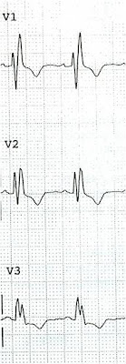
 Here is what V1-V3 should look like in RBBB:
Notice inverted T-waves, discordant to the majority of the QRS (the R' wave is the majority) and a small amount of discordant ST depression

He continued to have severe pain, so more ECGs were recorded, at 0927 and 0942:

There is increasing ST elevation in inferior leads without ST depression in aVL, and dynamic T-waves in V2 - V4.

Not much change from 15 minute prior, but abnormalities are persistent.

Here are V1-V3 from the last 3 ECGs:


It is now clear that  there is cardiac ischemia.  Inferior?  Anterior? Both?  Wraparound?


The patient was very hypertensive and had back pain, so they did a CT of his chest to rule out dissection.  It was negative. 

Because of the dynamic T-waves and crushing chest pain, he went directly to the cath lab where they found an open wraparound LAD with a large dangerous thrombus.

The peak troponin I was only 10 ng/mL and the patient did well.

Serial ECGs and close attention saved his anterior wall.

Here is the post PCI ECG:
T-waves are still upright.


Tuesday, April 24, 2012

Cardiac Ultrasound may be a surprisingly easy way to help make the diagnosis


This case was contributed by Vince DiGiulio.

A 63 year old woman who was otherwise healthy had a syncopal episode. She had no CP, no SOB, but did have dyspnea on exertion. Her BP was 106/56 with a pulse of 95. Here is her ECG:
There is sinus rhythm at a rate of 95 (tachycardic, depending on your definition of 90 vs. 100).  The QRS was measured by the computer at 117 ms (but to my eye is normal)  There is an rSr' in V1, and if the QRS really is long, this is an incomplete RBBB (I don't think it is long).  Computerized QTc is 435 ms.  There is abnormal T-wave inversion in leads V2-V5, suggestive of ischemia.  There is another finding which suggests the diagnosis.


The ECG differential includes Wellens' syndrome and pulmonary embolism.

Here is her previous ECG from 2 months prior:
There is sinus rhythm at a normal rate (75).  There is abnormal T-wave inversion in lead V2 only now, but the S1Q3T3 (the finding which suggests PE) is here also. 

So is this just baseline T-wave inversion?  Wellens'?  Pulmonary embolism?  First, the increased heart rate should always sway you towards PE.  Wellens' syndrome implies open arteries, perfusion of the myocardium, and the patient uncommonly has tachycardia.  Baseline T-wave inversion is an unlikely explanation because there is extension out to V5.  PE is very likely with this ECG.  

An EMT student who has never had any ultrasound training, who like this blog, likes EKGs, and likes to experiment with ultrasound was working in this ED where the attending emergency physicians do not use ultrasound frequently.  He had learned ultrasound from online sources only, including our own hqmeded, and related sites. 


The thick white arrow shows the right ventricle, which is dilated.  The thin red arrow shows the left ventricle, which should not be smaller than the RV, as it is here.


This self-trained EMT rolled the ultrasound into the patient's room and found this very large RV.  The patient underwent immediate CTPA, had a submassive saddle embolus, and underwent immediate catheter directed thrombolysis. 

My colleague Dave Plummer points out that: 

To be clear, the bedside ultrasound shows indirect evidence of PE by RV dilation only in cases of acute cor pulmonale.  This will manifest as:
1) dilated RV  
)2 hypo kinetic RV and 
3) thin walled RV.    

This is common to any acute RV outflow obstructions, including valvular.  As you point out, it can be discerned with minimal training

The ECG in PE:  

The ECG is not sensitive for PE, but when there are findings such as S1Q3T3 or anterior T-wave inversions, or new RBBB, then they have a (+) likelihood ratio and the S1Q3T3, or even just the T3, may help to differentiate Wellens' from PE. 

Stein et al. found normal angiograms in only 3 of 50 patients with massive PE, and 9 of 40 with submassive PE.  Today, however, that number would be lower because we diagnose more of the submassive PEs that have minimal symptoms.

This is a paper worth reading: Marchik et al. studied ECG findings of PE in 6049 patients, 354 of whom had PE.  They found that S1Q3T3 had a Positive Likelihood Ratio of 3.7, inverted T-waves in V1 and V2, 1.8; inverted T-waves in V1-V3, 2.6; inverted T-waves in V1-V4, 3.7; incomplete RBBB 1.7 and tachycardia, 1.8. Finally, they found that S1Q3T3, precordial T-wave inversions V1-V4, and tachycardia were independent predictors of PE. 

What is an S1Q3T3?  Very few studies define S1Q3T3.  What is it?   It was described way back in 1935 and both S1 and Q3 were defined as 1.5 mm (0.15 mV).  In the Marchik article, (assuming they defined it the same way, and the methods do not specify this), S1Q3T3 was found in 8.5% of patients with PE and 3.3% of patients without PE.

Kosuge et al. showed that, when T-waves are inverted in precordial leads, if they are also inverted in lead III and V1, then pulmonary embolism is far more likely than ACS.  In this study, (quote) "negative T waves in leads III and V1 were observed in only 1% of patients with ACS compared with 88% of patients with APE (p less than 0.001). The sensitivity, specificity, positive predictive value, and negative predictive value of this finding for the diagnosis of PE were 88%, 99%, 97%, and 95%, respectively. In conclusion, the presence of negative T waves in both leads III and V1 allows PE to be differentiated simply but accurately from ACS in patients with negative T waves in the precordial leads."

Witting et al. looked at consecutive patients with PE, ACS, or neither. They found that only 11% of PE had 1 mm T-wave inversions in both lead III and lead V1, vs. 4.6% of controls.  This does not contradict the conclusions of Kosuge et al. that when there are T-wave inversions in the right precordial leads and in lead III, PE may indeed by more common.  In my experience, this is true, but needs validation in a study of similar methodology. Supporting Kosuge, Ferrari found that anterior T-wave inversions were the most common ECG finding in massive PE. 


Thursday, April 19, 2012

Is This a Simple Right Bundle Branch Block?

A 72 year old male presented in shock.  Here is his initial ECG:
There is a wide complex at 194 ms, with RBBB. Anything else?


Here is his baseline ECG, also with RBBB:
There is sinus tach with otherwise normal RBBB; the QRS is 129 ms, average for RBBB.


The physicians appropriately recognized that the QRS in the first ECG was far too wide, and that there were peaked T-waves.  They immediately administered Calcium, Bicarbonate, D50 and insulin, and repeated the ECG:
The QRS is 164 ms (still long for RBBB) and the  T-waves are less peaked.

The initial K returned at 7.9 mEq/L, and the repeat K was 5.1 mEq/L (which seems like a rather impossible improvement in a short amount of time, but there is certainly some significant improvement on the ECG).

The patient had urosepsis with renal failure, was resuscitated and dialyzed.

This was recorded the next day, with the K at 3.3 mEq/L:


RBBB by definition has a long QRS (> 120 ms).  But very few are > 190 ms.  Literature on this is somewhat hard to find, but in this study of patients with RBBB and Acute MI, only 2% of patients with pre-existing RBBB had a QRS duration > 200 ms.  This study only reported durations in 10 ms intervals up to 150 ms, but one might extrapolate from it that approximately 10% of patients with baseline RBBB have a QRS duration > 160 ms.  194 ms would be quite unusual.

The point of this is that if you see BBB with a very long QRS, you must suspect hyperkalemia.  Then of course the peaked T-waves should tip you off.   Unless a patient has severe hypercalcemia (this should be evident by a short QT on the ECG as seen at the bottom of this post), or severe hyperphosphatemia (which is very unusual), treatment with calcium is harmless if you read an ECG falsely positive for hyperkalemia.

So don't wait for the laboratory K or you might be resuscitating a cardiac arrest (see the case with ECGs #3 and #4 of this post).

How about LBBB, asks one reader?

In this study of consecutive patients with LBBB who were hospitalized and had an echocardiogram, 13% had a QRS duration greater than 170 ms, and only 1% had a duration greater than 190 ms.


Sunday, April 15, 2012

Pseudonormalization of T-waves: STEMI

Here are other cases of pseudonormalization.

A 61 y.o. female with no h/o coronary disease presented with 3 days of nausea, vomiting, 3-4 liquid stools, and shortness of breath, especially with exertion. She denies fevers, chills, cough, chest pain, chest pressure, chest heaviness, diarrhea and abdominal pain. Her risk factors are HTN, smoking, and hyperlipidemia. 

Here is her initial ECG at 0508:
Sinus rhythm.  There are Q-waves in III and aVF diagnostic of old MI, and with the T-wave inversion in these leads, it appears to be fairly recent.  The inverted T-waves and relatively small Q-waves suggest that this was infarction was reperfused before it was complete.  T-wave inversion is often due to reperfusion and are thus sometimes called "reperfusion T-waves."  There is also ST depression in I, II, V4-V6 very suggestive of ischemia.

The Na returned at 117 mEq/L, but the troponin I returned at 8 ng/ml.   So the clinicians repeated the ECG at 0742:
There is now minimal ST elevation in III and aVF, and even more important, the T-waves have become upright in III and aVF, (although biphasic), with reciprocal ST segment and T-waves in I and aVL.  This is classic "pseudonormalization" of T-waves. 

Pseudonormalization of T-waves: "Normalization" because upright T-waves are usually normal, but "Pseudo" because even though they are upright, the fact that they are upright in this case is indicative of coronary occlusion.

She was moved to the critical care area, and at 0810, this ECG was recorded:
This is like the first one.  The involved artery has now reperfused, so the T-waves are inverted again ("reperfusion T-waves.")  so one would expect the artery to be open.  If this were a patient with chest pain, the pain would likely be resolved.  These T-waves are the analog to Wellens' T-waves which are described for the LAD or anterior wall.
 
The initial troponin I was 8 ng/ml, confirming that the infarct seen on the first ECG was recent.  The patient went for immediate PCI of a 95% distal RCA with hazy segment due to thrombosis.

Here is the ECG after PCI:

Not much changed from the previous.


Troponin I peaked at 18.  The inferior wall was akinetic on echo.

Learning points:

1.  Beware dyspnea in elderly women especially
2.  Subacute MI may present with T-wave inversion with or without Q-waves
3.  Subacute MI may have re-infarction
4.  If  T-waves of subacute MI turn upright (pseudonormalization), this is indicative of re-occlusion of the infarct-related artery

Monday, April 9, 2012

Repost: what is the diagnosis?

I'm reposting an edited version of this post from June 2009.

This is a 46 yo male with acute onset of chest pain, in distress, who called the ambulance. He arrived and had this ECG recorded at 0118 AM. 

There is 1 mm of ST elevation in V2 and V3, so this meets the criteria for reperfusion by the ACC/AHA guidelines. Unfortunately, the majority of patients who meet such "criteria" do not have MI. The most common reason for ST elevation is early repolarization.

This could be early repolarization or acute anterior STEMI.  How do we tell the difference?

A followup ECG and the answer is below.


In this case, the limb leads are suggestive of ischemia, with some subtle ST depression inferiorly, suggesting pending ST elevation in aVL due to lateral MI from proximal LAD occlusion.

If we apply the early repol vs. acute MI rule [see excel spreadsheet down the right side of the blog, which uses the formula (1.196 x STE at 60 ms after the J-point in V3 in mm) + (0.059 x computerized QTc) - (0.326 x R-wave Amplitude in V4 in mm)], and enter these values: STE at 60 ms after the J-point is 3.0 mm, QTc is 420 ms, and R-wave amplitude in V4 is 1.5 mm), then we get a result of 27.9 (it is anterior STEMI if the value is greater than 23.4). 

A simple rule is the R-wave rule, which depends on the fact that, in BER, the R-wave is always well developed: If the mean R-wave amplitude from V2-V4 is less than 5 mm, then it is almost certainly MI. If greater than 5 mm, it is probably BER. A cutoff of 5 mm gives a sensitivity for MI of about 70%, but a specificity of greater than 95%.  This ECG has very poor R-wave progression and so it cannot be early repolarization.

The rule relies on the findings that the mean ST elevation was higher in the MI group, and the mean QTc in BER is shorter (mean = 390 ms vs. 426 ms), and mean R-wave amplitude is greater.

The clinicians were suspicious of MI, so they were smart to obtain serial ECGs. They obtained the second ECG at 0143:

This shows unequivocal straightening of the ST segments, compared to the first ECG. This ST straightening results in T waves which are fattened and "hyperacute". This is diagnostic of anterior STEMI.

In case you can't see this difference in the straightening of the ST segment, here they are side-by-side:


The diagnosis was not made until more ECGs were recorded, so reperfusion was slightly delayed.  He had a 100% proximal thrombotic LAD occlusion with TIMI-0 flow. It was opened and stented.  The peak troponin was 138 ng/ml, so this was a large AMI.


Friday, April 6, 2012

Inferior hyperacute T-waves and ST elevation. Angiogram is normal. What happened?

I recently received this from a reader:

"I recently saw this 31 year old male with central chest tightness that started at 530am and lasted 30 min.  He came to ED pain free at 7 AM with no other symptoms.  The initial ECG showed J point elevation in inferior leads.  The morning doctors decided to do serial cardiac enzymes.  The first trop I was negative and subsequently went up slightly.  Patient was managed as NSTEMI.  A few days later the coronary angiogram was completely normal.   Please advise me on this ECG.  Should we have done a bed side echo to look for wall motion abnormality and would that have made a difference in management?"
See interpretation below


Here was my response:

"This is diagnostic of inferior injury (STEMI).  There is inferior STE with hyperacute T waves, reciprocal ST depression and T-wave inversion in aVL. Also STE in V5 and V6. It spontaneously reperfused. A negative cath does not prove anything.   There was either thrombus that completely lysed or coronary spasm.  Did they do IVUS (intravascular ultrasound of coronaries)? That would identify extra coronary plaque as the source of ACS."

A high quality echocardiogram would most likely have shown a wall motion abnormality.

"If you can find a repeat ECG, it will show resolution of the findings on subsequent ecg's. Can you find one?"

He did find one and here it is:
Complete resolution of ST elevation


The troponin I peaked at 5.  The angiogram was completely normal. The patient was treated with clopidogrel.

This was a reperfused STEMI.

Normal Coronaries in Suspected Acute Coronary Syndrome:

The 4 articles below, about 8% of cases referred for primary angioplasty of STEMI have completely normal coronaries.  Many of these are false positive ECGs, but some are MI with due to spontaneous reperfusion.  If there is spontaneous reperfusion, the ECG will always show typical evolution or resolution.  

This case has an unequivocal ECG; it is clearly STEMI.  In cases in which the ECG is not unequivocal, absence of change over time proves it is not a STEMI.

By the way:
1) Non STEMI but with ST elevation due to non-AMI etiologies (early repol, LVH, etc.) is never documented in these studies.  
2) Furthermore, the mere presence of non-obstructive coronary disease, unless one sees a culprit lesion, does not prove that the symptoms were due to AMI.  

Whether there is ACS or not, troponin will often be positive because troponin is nonspecific: it is elevated is many states including pulmonary embolism, myocarditis, stress cardiomyopathy, dilated cardiomyopathy, hypertensive cardiomyopathy, and more.  This has recently been called a "type II MI". 

Coronaries can now be evaluated with other means, such as intravascular ultrasound (IVUS).  Even when there is stenosis, most atherosclerosis is extraluminal.  It can be seen with IVUS, but not with a "lumenogram," which is what an angiogram is.  These atherosclerotic plaques can cause intralumenal thrombosis with STEMI.  If the clot lyses completely, the ischemia resolves and the angiogram is normal.


Bibliography, with edited abstracts


There were 821 cath lab activations and 86% were treated by mechanical revascularization. In 76 patients (8.5%), no coronary artery stenosis was documented. Observations documented angiographically included coronary spasms (6.6%) and muscle bridges (5.3%). During a mean follow-up of 11.2±6.4 months, one patient developed an acute myocardial infarction requiring coronary intervention. All other patients were free of any cardiac event.


Article 2, full text


Of 898 patients who had cath lab activations for primary PCI, normal coronary angiograms were obtained for 26 patients (2.6%). Among these, the diagnosis at discharge was a small myocardial infarction in seven patients (0.7%), acute (peri)myocarditis in five patients, dilated cardiomyopathy in four patients, hypertension with left ventricular hypertrophy in three patients, pulmonary embolism in two patients and misinterpretation of the electrocardiogram (ie, no cardiac disease) in five patients. Seven patients with small infarctions underwent angiography within 30 min to 90 min of complete relief of the signs of acute ischemia, and thus, angiograms during pain were not taken.   None of the 898 patients catheterized during ongoing symptoms of ischemia had a normal coronary angiogram. Spontaneous coronary spasm as the only cause (without underlying coronary atherosclerosis) for the evolving infarction was not seen. Thus, the causes of the seven small infarcts in patients with normal angiograms remain uncertain.


Characteristics of 690 consecutive patients with presumed STEMI referred for primary PCI.  87 (13%) had angiographically normal coronary arteries and were compared with patients with angiographically shown culprit lesions (control group; n = 594). Nine patients with significant coronary disease, but no identifiable culprit lesion, were excluded. Electrocardiograms (ECGs) from both groups were reviewed by 2 cardiologists blinded to angiographic findings.  On expert review of ECGs, 55% of patients in the normal coronaries group had ST-elevation criteria for STEMI (vs 93% in the control group, but the ECG was considered consistent with a diagnosis of STEMI by both observers in only 33% (vs 92% in the control group)   Left branch bundle block independently correlated with normal coronary arteries on multivariate analysis (odds ratio for STEMI 0.016).   The discharge diagnosis in the normal coronaries group was predominantly pericarditis (n = 72; 83%), but these were not adjudicated by the authors.  Other diagnoses were myocarditis in 3 patients (3%), Takotsubo cardiomyopathy in 2 patients (2%), presumed coronary spasm secondary to intravenous drug abuse in 2 patients (2%), cryptogenic AMI in 1 patient (1%), dilated cardiomyopathy in 1 patient (1%), massive pulmonary embolus in 1 patient (1%), cholelithiasis in 1 patient (1%), and pneumonia in 1 patient (1%).

The most likely alternative diagnosis suggested by both observers for the non-AMI ECGs in the normal coronaries group was normal variant ST changes (25% observer 1 and 26% observer 2) and early repolarization abnormality (25% observer 1 and 14% observer 2). 


The medical records of 941 patients undergoing coronary arteriography for presumed ACS within 48 h of onset were critically reviewed. In 70 patients (7.4%, 35 males) no CAD was documented. Alternative substrates of acute myocardial ischemia included coronary artery anomalies (7 patients, 10%), coronary spasm (10 patients, 14.3%), spontaneous coronary dissection (2 patients, 2.8%), paradoxical embolism through a patent foramen ovale (4 patients, 5.7%), embolism from left atrium or calcified aortic valve (4 patients, 5.7%), imbalance between oxygen demand and supply (20 patients, 28.5%), mitral valve prolapse (11 patients, 15.7%). No alternative substrates were found in 12 patients (17.1%). Absence of CAD is an uncommon finding in patients undergoing coronary artery angiography for ACS.


Monday, April 2, 2012

Intermittent third degree heart block due to stuttering inferior STEMI

A 71 year old male complained of near syncope and SOB without any chest pain.  His pulse was slow.  He was put on the monitor.  Here is the rhythm strip:


There is 3rd degree AV block at a rate of 48

A 12 lead ECG was recorded about 5 minutes later:

Sinus rhythm at a rate of 60, with a normal PR interval, and right bundle branch block with no ischemia.  There are inferior Q-waves diagnostic of old inferior MI.  The upright T-waves in V1-V3 are slightly unusual for RBBB, but nondiagnostic.  It is not known whether RBBB is new or old.

21 minutes later the patient became bradycardic again, so this ECG was recorded:

There is 3rd degree AV block at a rate of 43 (again), still with RBBB but now with an obvious inferior STEMI.  There is also some ischemic ST depression in V5 and V6.  The sinus node has a rate of about 75, so there is no excess vagal tone, and therefore no utility of atropine.

One might think that because there is associated RBBB, that it is infranodal AV block.  But the RBBB was present when the block and ST elevation were not, so it is likely a baseline feature of this patient's ECG.  Furthermore, inferior STEMI is much less likely to cause infranodal block.  The blood supply to the AV node is from the RCA, and the infranodal (HIS bundle) area is supplied by the LAD.  Therefore, intranodal (in the AV node) block is much more likely than infranodal (below the AV node, in the bundle of HIS).

When in the node, AV block has a much better prognosis than when below the node.    

In the setting of STEMI, the ACC/AHA guidelines, page 618, recommend permanent pacing as a Class I indication for:

1. persistent second-degree AV block in the His-Purkinje system (infranodal) with bilateral bundle-branch block or third-degree AV block within or below the His-Purkinje system after STEMI. (Level of Evidence: B)

2. transient advanced second- or third-degree infranodal AV block and associated bundle-branch block. If the site of block is uncertain, an electrophysiological study may be necessary. (Level of Evidence: B)

3. persistent and symptomatic second- or third-degree AV block. (Level of Evidence: C)

Persistent here is important, because it is likely that, after opening the artery, the block will resolve, and thus will not be persistent.

The patient was comfortable while supine, even with 3rd degree AV block.  The block was intranodal.  The patient would soon have PCI.  So no pacing was necessary.  One should have external (transcutaneous) pacing at the ready in case of worsening block, but with inferior STEMI this is unlikely to be necessary.

He went to PCI and had a mid-RCA occlusion opened.  Here is his post PCI ECG:


Sinus rhythm without any AV block.  ST elevation is resolved and there is inferior T-wave inversion (reperfusion T-waves).  RBBB is persistent, the T-waves and ST segments are more normal for RBBB.  Is this normalization after resolution of ischemia?  Or is this T-wave inversion due to reperfused posterior MI?  Without a previous ECG, it is hard to know for sure.
 

Initial troponin I was 0.035 ng/ml (99% reference, 0.034) and peaked at 13.4.  Echo showed infero-postero-lateral wall motion abnormality.

Learning points:

1. STEMI does not always have chest pain or SOB.  It is wise to obtain serial ECGs when there are ischemic symptoms, and especially with AV block.

2. Inferior STEMI may cause AV block, but it is intranodal

3. Infranodal AV block, especially in the context of anterior STEMI, is very dangerous and will probably require internal pacing unless the artery can rapidly be opened. Before the artery is opened, it is at least likely to require external pacing.