Monday, November 22, 2010
Tuesday, November 16, 2010
Wide Complex Tachycardia; It's really sinus, RBBB + LAFB, and massive ST elevation
these are all a mix of ventricular tachycardia and SVT with aberrancy.
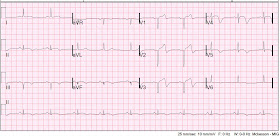
This 51 yo male complained of chest pain, then had a v fib arrest. He was resuscitated and brought to the ED where this ECG was recorded. He was in cardiogenic shock.
There is tachycardia, and there is a wide complex. This wide complex tachycardia could easily be misdiagnosed as V tach. However, there are p-waves, and this is a classic RBBB + LAFB (left anterior fascicular block) morphology. When V tach originates in the left ventricle, there may be an RBBB-like complex, but because VT originates in the myocardium, not in the left bundle (as does RBBB), it does not look exactly like RBBB, as this one does. The left anterior fascicular block can be diagnosed by the left axis deviation. RBBB alone would have S-waves in I and aVL; since there are late large R-waves, there is LAFB.
So now we can say it is sinus tach with RBBB + LAFB.
Is there ST elevation? One must find the end of the QRS in order to determine this. I have done this and marked it up in the image below. The end of the QRS is easy to find in V1. One can then draw a line down to the rhythm strip at the bottom, which is lead II. Thus, you can find where in lead II is the end of the QRS. Then you can go to all parts of the ECG to find the end of the QRS.
As you will see, this results in the discovery of ST elevation in V2-V4 and I and aVL, diagnostic of anterolateral STEMI.
The ED providers activated the cath lab, but the interventionalist refused to come in because it was "not a STEMI". The patient died 8 hours later of cardiogenic shock.
Tuesday, November 9, 2010
Sunday, November 7, 2010
Pure Posterior STEMI, not a rare event

There is ST depression with upright T-wave in leads V2-V6, maximal in V3. There is no ST elevation anywhere on the ECG. There are those who deny the existence of posterior STEMI; they argue that coronary anatomy makes it "extremely unlikely." But fact has a way of overturning theory.
After therapy with IV nitroglycerine and an aspirin, the ST depression and chest pain resolved. Because of issues with some contraindications to antiplatelet and anticoagulation therapy, and because the ECG and symptoms had resolved, he was not taken immediately for cath. He did have an echo confirming a new posterolateral wall motion abnormality.
Next day cath showed a 100% Ramus intermedius occlusion; it was opened and stented. Max TnI was 14 ng/ml. Echo showed corresponding new WMA and EF was 54%.
There are many studies that indirectly reveal that the percent of STEMIs that are isolated posterior is between 3 and 11% (about 8%). More recently, a substudy of the recent TRITON-TIMI 38 trial comparing Prasugrel to Clopidogrel for ACS enrolled 13,608 patients; 1198 had isolated ST depression in V1-V6. Of these, 314 (26%) had occlusion (TIMI 0 or 1 flow) of the infarct-related artery (i.e., STEMI).
There were 3534 other STEMIs in this study, not including the 314 with ST depression only (posterior STEMI). Add these 314 to the 3534 and you have 314/3848 (8.1%) of STEMI have pure isolated posterior STEMI. This conforms with the previous smaller studies. Moreover, the cath was done a median of 29.4 hours after presentation, so this does not account for those arteries that spontaneously reperfused (about 25% of STEMI will reperfuse with antiplatelet and antithrombotic therapy alone within one day -- old data). Thus, there were probably even more occluded arteries.
Only 14/314 (4.5%) were interpreted by the investigator as STEMI. None of the patients with an occluded artery had an ECG to PCI time <6 hours.
This is not a "rare" event.
Saturday, November 6, 2010
Subacute STEMI Masked by a Wide Complex
Case:
This 46 yo male with no h/o MI or coronary disease presented with 2 days of palpitations, nausea and dizziness and intermittent chest pain that started while walking. The chest pain was never prolonged and constant. Here is his initial ECG (816 AM):
V1-V3 have RBBB morphology, but the initial r of the rSR' is replaced by a Q-wave. V3 has an RBBB pattern with ST elevation. There is 1 mm of ST elevation in V1-V3 in the presence of RBBB; this is abnormal, but when there is a Q-wave, it can be due to old MI with persistent ST elevation. ST segments in RBBB in V2 and V3 are usually negative, opposite the tall R' wave. Any ST elevation is abnormal.
This was unrecognized, and at 941 AM another ECG was recorded:
The troponin peaked at 175, there was a large anterior, septal and apical WMA with EF of 40%.
Here is a slightly later recording:
 |
| The escape rhythm with RBBB morphology remains, and all T-wave changes are obscured. Thanks to VinceD for recognizing the retrograde (inverted) p-waves buried in each RBBB complex. |
2) In RBBB, any ST elevation in V1-V3 is abnormal


