Wednesday, May 8, 2024

A young man with palpitations.

A 30-something presented with chest pain, palpitations, and SOB.  He has had similar symptoms for 4 years, but has never been evaluated.

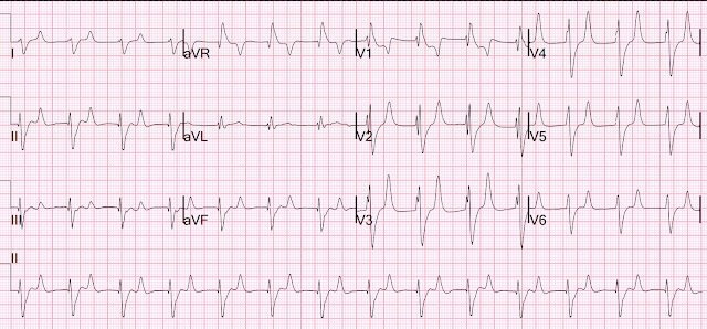
Here is his presenting ECG, which was sent to me real time, along with the 2nd ECG below:

Regular Narrow Fast without P-waves.  PSVT.  
It is very difficult to tell if this is:
 1) AVNRT or 
2) orthodromic AVRT 
(Orthodromic AVRT = WPW with orthograde conduction down the AV node and retrograde up an accessory pathway)
See Ken Grauer's discussion below in differentiating AVNRT from orthodromic AVRT.


Shortly after arrival, the patient spontaneously converted to this rhythm (also sent to me, along with the first):

What do you think?


The two ECGs above were texted to me with the text: "Young Guy came in in SVT but now in and out of irregular wide complex tachycardia. -- not sure if polymorphic VT vs. a fib with WPW."

My response: "Definitely not polymorphic VT.  Definitely atrial fibrillation.  Probably WPW but is very slow for atrial fib withWPW.  I would just cardiovert electrically."

Smith: the rhythm is irregularly irregular without P-waves.  There are delta waves and polymorphic QRS (but NOT polymorphic VT!).  So this looks like WPW with Atrial fibrillation.  What is unusual is that the rate is not REALLY fast, as you expect when there is atrial fib with an accessory pathway.  The shortest R-R inteval that I see is between the 6th and 7th beat and is 280 ms, which is not dangerously short.

Nevertheless, you NEVER want to give an AV nodal blocker to Atrial fib with WPW:

1. The reason to avoid AV blockers in WPW is that, in addition to blocking AVN, they actually enhance conduction via AP by shortening refractory period, which can produce VF.

2. Adenosine is considered safe for termination of AVRT in WPW, but the pro-arrhythmic effects of adenosine can rarely cause AF, so do not give AVN blocker to WPW unless you have defib pads placed.

The essential features of A Fib WPW are:

1. Irregularly irregular

2. Polymorphic

3. Wide

4. Some very short R-R intervals

Never give an AV nodal blocker to Atrial Fibrillation with WPW; it can result in ventricular fibrillation.  This means NO calcium channel blocker, beta blocker, adenosine, or digoxin.

Cardioversion can be done pharmacologically (usually procainamide), but why would you want to do that?  Only if you are unable to perform procedural sedation or if the patient refuses.

So electrical cardioversion was done (using etomidate sedation.  I prefer low dose propofol, just enough for amnesia, but not enough to result in serious hypotension).

Here is the post cardioversion ECG:

Definite pre-excitation (delta waves due to accessory pathway, "bypass tract").  Pre-excitation results in a short PR interval.


Electrophysiology note:  "In the context of pre-excited atrial fibrillation, we would recommend proceeding with mapping and ablation of accessory pathway (particularly given high risk features including his shortest pre-excited R-R interval is < 250 ms). It does appear likely that his pathway may anteroseptal, which does increase risk of damage to AV node."

More explanation from electrophysiology:

"ECG only records 10-seconds worth of data, therefore long rhythm strip is essential to make sure ample data is available. Fortunately in the stabilization room, they had recorded a long rhythm strip (Smith: not seen by me and not shown) during atrial fibrillation and some R-R intervals were very short approaching 250 msec."

He was taken for immediate ablation that day in the EP lab, but (explanation from electrophysiology) this ECG was recorded:

No pre-excitation, therefore, no ablation attempted


Electrophysiolgy explains: "These accessory pathways can be very superficial fibers and during the first procedure, it got mechanically “bumped” before turning on ablation and therefore NO ABLATION WAS PERFORMED in absence of preexcitation, given high risk location. Despite of waiting for an hour pathway conduction did not come back (but they do invariably come back after such mechanical bump). Therefore repeat procedure was performed with Cryo-ablation after the accessory pathway conduction had returned."


The next morning, this ECG was recorded:

What do you think?


The delta waves are back.  There is recurrent pre-excitation.


The Ablation was done.


He was taken for another ablation and this ECG was recorded:

Normalized.
He was discharged.


===================================

MY Comment, by KEN GRAUER, MD (5/8/2024):

===================================
I found today's case extremely interesting regarding a number of points about reentry SVT rhythms, with or without AP (Accessory Pathway) participation. I'll add the following points to Dr. Smith's excellent discussion.
  • For clarity in Figure-1 — I've labeled the first 2 tracings in today's case.

Figure-1: I've labeled the first 2 tracings in today's case.

Can We Distinguish between AVNRT vs Orthodromic AVRT?
As per Dr. Smith — the principal differential diagnosis between the regular SVT reentry rhythms, is between AVNRT (AtrioVentricular Nodal Reentry Tachycardia — in which the reentry circuit is contained within the AV Node) — vs orthodromic AVRT (AtrioVentricular Reciprocating Tachycardia — in which the reentry circular first travels down the normal AV nodal pathway, and then conducts back to the atria over an Accessory Pathway).
  • As I illustrated in My Comment at the bottom of the page in the March 6, 2020 post of Dr. Smith's ECG Blog — while not infallible, the finding of a very short RP' interval suggests the mechanism of the reentry SVT rhythm is AVNRT, in which the impulse travels back to the atria over the "fast" AV nodal pathway. This is because when the reentry circuit is entirely contained within the AV node — it takes minimal time to return to the atria.

  • In contrast — the finding of a moderately long RP' interval suggests that the mechanism of the reentry SVT rhythm is orthodromic AVRT, in which a "concealed" AP may be participating in the reentry pathway. Because the AP lies outside of the AV node — the time to circulate around the reentry pathway and conduct back to the atria (retrograde) is longer than when the entire reentry circuit is contained within the AV node.

As I illustrate in the March 6, 2020 post — retrograde conduction can often be seen during a reentry SVT rhythm! 
  • IF you look for retrograde P wave conduction during a regular SVT rhythm — You'll begin to find it much more often than you might have imagined! On occasion — recognizing retrograde conduction may provide a KEY clue to the mechanism of a tachycardia.
  • To LOOK for retrograde P waves:  i) Look in the inferior leads for a small negative notch that occurs at the very end of the QRS, or fairly soon thereafter; orii) Look for a small positive notch in lead aVR and/or lead aVL and/or lead V1andiii) Confirm that the notching you identified during the regular SVT rhythm truly reflects retrograde atrial activity (rather than being a part of the QRS or the result of artifact) — by noting that this notching is no longer present after conversion to sinus rhythm. (My Figure-2 in the March 6, 2020 post illustrates this confirmation).

Regarding Today's CASE: 
Unfortunately — I found the initial ECG in today's case equivocal with regard to my search for retrograde P waves:
  • As shown in Figure-1 — the rhythm in today's initial ECG is a regular SVT (ie, Narrow-complex tachycardia) at ~190/minute, without sinus P waves.
  • I initially thought the small negative notch at the very end of the QRS in lead II — and the small positive notch at the very end of the QRS in lead V1 — both represented retrograde P waves with a very short RP' interval (within the RED circles in these leads). If so — this would strongly suggest AVNRT of the "slow-fast" type (which is by far the most common form of AVNRT) as the mechanism of today's initial rhythm.
  • That said — I thought the more-pointed-than-expected negative deflections in leads III and aVF — and the pointed tip of the upright T wave in lead aVL — might represent retrograde P waves, in which case the RP' interval would instead be moderately long (within the BLUE circles in these leads).

  • BOTTOM Line: Given conflicting findings regarding my search for potential retrograde atrial activity — I could not distinguish between AVNRT vs orthodromic AVRT on the basis of ECG #1 alone. Sometimes you can distinguish between AVNRT vs orthodromic AVRT — but not in today's initial tracing.

WHY CARE if the Mechanism is AVNRT vs Orthodromic AVRT?
While emphasizing that overall initial management of AVNRT and orthodromic AVRT are similar — there are some differences to be aware of that may have a role in treatment decision-making.
  • WPW is recognized on ECG by the presence of 3 Findingsi) A short PR interval; ii) Delta waves in a number of leads; andiii) QRS widening. 
  • It's important to appreciate that patients with an AP may not always manifest these 3 findings that are characteristic of complete or predominant preexcitation. Instead — Patients with an AP may at times manifest partial or even no preexcitation (with the relative percentage of impulses traveling over the AP vs the normal AV nodal pathway being highly variable). IF the relative amount of preexcitation at any given time is minimal — then delta waves, PR interval shortening, and QRS widening may be barely (if at all) detectable.

  • APs may conduct forward (anterograde) and/or backward (retrograde). While APs are usually capable of conducting in both directions — forward or backward conduction may not be possible in some patients. This leads to the concept of a "concealedAP — in which only retrograde conduction is possible over the AP. 
  • Up to 15% of patients with a regular reentry SVT rhythm who are referred for catheter ablation may have a "concealed" AP, in which delta waves are never seen because forward (anterograde) conduction over their AP is not possible (Pappone and Santinelli — ESC: July, 2018)

  • KEY Point: Synthesizing the concepts in the above bullets means that a significant percentage of the time when treating a new patient with a regular reentry SVT rhythm, but without clear sign of atrial activity — that despite never seeing delta waves, the patient may have a concealed AP (ie, that rather than AVNRT — the regular SVT may be orthodromic AVRT).
  • The "good" news — is that initial treatment of such patients with a regular reentry SVT rhythm is the same (ie, Adenosine, Verapamil/Diltiazem, ß-Blocker) — because AV nodal blocking agents will successfully interrupt the reentry circuit in both AVNRT and orthodromic AVRT.
  • The "less good" news — is that in up to 1/3 of patients with orthodromic AVRT — spontaneous AFib may develop at some point — presumably predisposed by "triggering" of AFib by AP-mediated reciprocating tachycardia (Ma et al — Exp Clin Cardiol 9(3): 196, 2004 — and Silverman et alin J Investig Med: Jan, 2018). And, as per Dr. Smith — use of AV nodal blocking agents (ie, Adenosine, Verapamil/Diltiazem, ß-Blockerare contraindicated in patients with AFib and WPW — because blockade of the normal AV nodal pathway may result in further acceleration of anterograde conduction over the AP, which may result in deterioration of the rhythm to VFib.
  • The other "less good" news — is at least the theoretical risk that using an AV nodal blocking agent to treat a regular SVT might precipitate AFib if the patient had a "concealed" AP (ie, if instead of AVNRT — the rhythm was orthodromic AVRT). Fortunately — precipitation of AFib by giving an AV nodal blocking agent to a reentry SVT that turns out to be orthodromic AVRT is rare (and consensus remains to treat reentry SVT rhythms with AV nodal blocking agents).


What About the Rhythm in ECG #2?
I was intrigued by the spontaneous development of the rhythm in ECG #2 — just a short while after ECG #1 without administration of any medication!
  • As per Dr. Smith — the rhythm in ECG #2 is irregularly irregular without P waves. While I hesitate in diagnosing delta waves in the absence of sinus P waves (because LBBB conduction may sometimes manifest initial QRS slurring) — the additional features of changing QRS morphology — and the finding of some very short R-R intervals alternating with some unexpectedly longer R-R intervals (ie, between beats #5-6) in this younger adult — suggested WPW with AFib (although definitive diagnosis of WPW was not made until the 3rd ECG was obtained, which showed return of sinus rhythm with short PR, delta waves and a similar-looking wide QRS).

  • PEARL #1: As we have highlighted in previous posts (See the March 11, 2020 post) — WPW can be immediately diagnosed when you see AFib with a wide QRS and an extremely rapid ventricular response (that at times attains a rate of between ~220-250/minute!).
  • Unlike PMVT (PolyMorphic Ventricular Tachycardia) in which QRS morphology usually changes dramatically from one beat to the next — the changing QRS morphology typically seen in WPW with AFib tends to be more subtle (due to modest variation in the relative percentage of preexcitation).

  • PEARL #2: Not all patients with WPW are at high risk of developing potentially life-threatening tachyarrhythmias. The risk of developing VFib during AFib in a patient with WPW is greatly increased when the SPERRI (Shortest Pre-Excited R-R Interval) measures below 220-250 msec. This corresponds to a shortest R-R interval that is barely more than one large box in duration. We do not see this in ECG #2 — as the shortest R-R interval is 7 little boxes in duration ( = 280 msec. — as I've labeled between beats #6-7).
  • How fast the ventricular response will be in association with AFib in a patient with WPW will depend on anterograde conduction properties of the AP. The overall rate of AFib in ECG #2 is ~150/minute (ie, There are 25 beats in the 10 second long lead II rhythm strip ==> 25 X6 = 150/minute). Since the ventricular response in ECG #2 is comparable to the rate range for any patient who develops new-onset AFib — definitive diagnosis of WPW was not made in today's case until the 3rd ECG was obtained.

FINAL Points in Today's CASE:  
  • Even though the SPERRI value during AFib in today's case was not below 250 msec. — catheter ablation was indicated because this younger man was very symptomatic with not just one, but two WPW-related tachyarrhythmias (orthodromic AVRT and AFib).
  • It is possible to have more than a single AP! (Goyal et al — StatPearls: July, 2023). While multiple APs in a given patient is not common — I initially wondered if that might be a possibility in today's patient, given the need for repeat ablation the next day. That said — the fact that morphology of the wide QRS was virtually identical in sinus rhythm after each ablation suggested this patient did not have more than a single AP (and in Dr. Smith’s discussion, under Electrophysiology Explains — we learn the reason the EP cardiology repeated the procedure the next day).
  • Finally — Did you notice the very tall T waves in multiple leads after each ablation? This finding is typical for a post-ablation memory T wave pattern — which is often considered evidence of a successful ablation (Silverman et al — J Investig Med: Jan, 2018).


Sunday, May 5, 2024

Do you need to be a trained health care professional to diagnose subtle OMI on the ECG?

An undergraduate (not yet in medical school) who works as an ED technician (records all EKGs, helps with procedures, takes vital signs) and who reads this blog regularly arrived at work and happened to glance down and see this previously recorded ECG on a table in the ED.  It was recorded at 0530:

What do you think?


The young ED tech immediately suspected LAD OMI.

He interprets here:

"This EKG is diagnostic of right bundle branch block and transmural ischemia of the anterior wall, most likely from an occlusion of the proximal LAD. There is a hyperacute distribution of T waves from V1 to V4. V1 has inappropriate ST elevation with a terminally negative T wave. The T waves in V2, V3, and V4 are symmetrical, upright, and too large for their preceding QRS complexes. Leads I and aVL have hyperacute T waves as well. There are reciprocal changes inferiorly, most pronounced in lead III with a downsloping ST segment followed by a terminally upright T wave."

_____________

Smith: RBBB with OMI often is difficult to diagnose on the ECG:

See what happens when the consultant is "Not convinced of STEMI"

______________

The ED tech knows the Queen of Hearts, and analyzed the ECG with the AI app:

OCCLUSION MI WITH HIGH CONFIDENCE

Register for access to Queen of Hearts here

The ED tech inquired and realized that the doctor had not appreciated the ECG diagnosis.  

The provider had sent the patient for an aortic dissection scan which had shown extremely heavy calcification of the LAD.

He learned more about the patient: A 77 year old female with a past medical history of hypertension and hyperlipidemia presented to the ED at around 0520 after waking up at 0400 with 10/10 chest heaviness radiating to both arms. 

The patient had continued to have chest pain.

Therefore, the provider had appropriately recorded another ECG (but unfortunately unnecessarily delayed by 45 minutes at 0615):

Now it is obvious to everyone, not only to an expert.

The cath lab was activated.

There was a 100% proximal LAD occlusion that was opened and stented.  

But 45 minutes later than it should have been.

Later, the ED tech found a previous ECG for comparison:

This further proves that the OMI findings were indeed OMI findings.

Measures of infarct size are not available, but it is certain that it would have been smaller with earlier intervention.

____________________________

Learning Points

Many of the most proficient interpreters of the ECG for OMI have little medical training.  But they have an interest in ECGs, lots of exposure to OMI ECGs as well as normals and mimics, an interest in acute coronary occlusion, and a talent for seeing the subtle waveform findings of acute OMI.  Such proficient interpreters include health care assistants and EKG technicians.  

Pendell Meyers had not started medical school by summer of 2012, but he had read every one of my blog posts over the preceding 4 years.  He was a paramedic at the time.  By the summer of 2012, he could read an ECG for OMI better than any doctor I knew.

A corollary to this is that though cardiologists have a vast knowledge of the heart, many just do not see OMI ECG findings that even an undergraduate might see.

Another corollary is that, due to the extreme difficulty and varying skill and talent at this, AI is the only answer.

And the Queen of Hearts interpreted OMI with High Confidence.

Register for access to Queen of Hearts here


Friday, May 3, 2024

Sudden shock with a Nasty looking ECG. What is it?

A 60-something woman complained of sudden severe abd pain. She was found by medics agitated, hypotensive, diaphoretic, and in shock.


There were 2 prehospital ECGs:

What do you think?


Smith: Uncertain supraventricular rhythm with PVCs. (See Ken Grauer's analysis below). There is "shark fin" in I and aVL, which is due to a combination of a large R-wave due to left anterior fascicular block plus downsloping ST elevation due to OMI.  There is reciprocal STD in inferior leads.  There is a rather large R-wave in lead V1 and a very large R-wave in V2, suggesting an atypical RBBB.  There is huge ST depression across the precordial leads.  There is STE in aVR.  Thus, there is high lateral OMI with diffuse ST depression.  

When I was shown this ECG, I said it looks like such widespread ischemia that is might be a left main occlusion, or LM ischemia plus circumflex occlusion (high lateral and posterior OMI).  Moreover, left main occlusion often presents near death.  In fact, most do not make it to the hospital alive, which explains why only a tiny percent of OMI are due to full LM occlusion.

Here is the Queen of Hearts interpretation:


There is a second prehospital ECG:

Again, supraventricular rhythm with RBBB and LAFB, shark fin, and STD maximal in V3.  Posterior and high lateral OMI.
But this time the Queen gets it wrong (thinks it is not OMI):


There were runs of VT:


Tha patient arrived in profound shock and had an ED ECG:

Now there is some evolution to include the ST elevation (rather than ST depression) in V4-V6.  This suggests that thrombus has propagated to occlude the LAD in addition to the circumflex.


It can be difficult to see ST elevation and depression when there is RBBB.  
--It helps to find the end of the QRS.  
--This is most easily done in lead V1.  
--Then you can draw a line down through leads V2 and V3 to the lead II rhythm strip across the bottom.  
--Then you can find the same location on the QRS in each 2.5 second interval.  
--Then you can draw the line up to each of leads I, II, III; then aVR, aVL, aVF; then V4, V5,V6.


The Queen of Hearts gets it right here:


The interventionalist stated that he could not do the procedure while the patient has a blood pressure of 45 systolic.

Therefore, the patient was put on arterio-venous ECMO.

Then an angiogram was done.

There was a 100% Left Main Occlusion (OMI).  It was opened and there was thrombus in the circ and LAD.

Unfortunately, the patient ultimately died.

Learning Points:

1. RBBB + LAFB in the setting of ACS is very bad.  Some patients have baseline RBBB with LAFB, but in patients with likely ACS, these are associated with severe infarction with cardiac arrest, cardiogenic shock or impending shock.

2. Patients with ACS and RBBB/LAFB usually have a left main vs. proximal LAD. 

Here are some cases of RBBB with LAFB: 

What is the Diagnosis in this 70-something with Chest Pain?


3. Left Main Non-Occlusive ACS presents with widespread ST Depression and STE in aVR.  Total Left Main Occlusion presents with different ECG findings which are multi-faceted.

See the variety of Left Main Occlusion ECGs here: 

How does Acute Total Left Main Coronary occlusion present on the ECG?


===================================

MY Comment, by KEN GRAUER, MD (5/3/2024):

===================================
Most patients with acute LMain Occlusion do not survive to make it to the hospital. Today's patient did make it to the hospital — but was in cardiogenic shock, and despite valiant attempt at treatment, succumbed soon after.
  • I focus my comment on some additional Learning Points to those highlighted by Dr. Smith.


ECG Findings with Acute LMain Occlusion:
As per Dr. Smith — the ECG findings of total acute LMain occlusion are multifaceted. In Figure-1 — I reproduce major points that I've summarized from Dr. Smith's August 9, 2019 post on the subject. The KEY Take Home Points are as follows:
  • There is no “single” ECG presentation for patients with acute LMain occlusion. Quite literally — You can see almost anything!
  • The reason for this highly variable ECG presentation, is that multiple territories may be involved to varying degrees — making it impossible to predict how much ST elevation you will see — and how much opposing (reciprocal) ST depression will attenuate (if not completely cancel out) these initial ST segment vector forces. 
  • The ST-T wave appearance in lead aVR can be anything when there is acute LMain occlusion.

Figure-1: Reasons for the varied ECG presentation of acute LMain occlusion — excerpted from Dr. Smith’s 8/9/2019 post (See text).


"Shark Fin" ST Elevation and Depression
As per Dr. Smith — the 3 12-lead ECGs in today's case all showed prominent "Shark Fin" ST segment deviations.
  • Dr. Smith illustrates in his discussion the delineation between the end of the QRS and the beginning of the ST segment for the 1st ECG done in the ED.
  • For clarity in Figure-2 — I illustrate this delineation point for ECG #1 (ie, the 1st EMS ECG) — whereby everything to the right of the vertical RED lines that I've drawn in ECG #1 represents either marked ST elevation (in leads I,aVL and aVR) — or marked ST depression (in leads II,III,aVF; V1,V2,V3; V5,V6).

NOTE: For those wanting more practice recognizing Shark Fin ST-T wave changes — we've shown cases of this entity in the following ECG Blog posts (among others): 


=====================================

What is the Rhythm in Today's Case?
Determination of the cardiac rhythm in today's case is of more than academic interest — since my initial "quick glance" of ECGs from this woman in her 60s who presented in shock — was that the QRS looked wide with an irregular rhythm that might represent a polymorphic VT.
  • Looking closer — I recognized the Shark Fin morphology (that I illustrate with the vertical RED lines in ECG #1 of Figure-2).

How then to approach the rhythm in ECG-1?
  • To Emphasize: I initially assessed the rhythm solely from ECG-1 — trying as I always to, "to put myself in the same position as providers in the field — who initially only saw ECG #1".

  • As Dr. Smith has noted — recognition of Shark Fin morphology told us that the seemingly wide and irregular rhythm in Figure-1 was almost certain to be supraventricular!

  • Despite the irregularity of QRS complexes — this rhythm is not AFib — because at least some definite P waves are present (RED arrows that I added at the bottom of ECG #1).
  • Given that there are at least some definite P waves — I looked more carefully for spots in which more subtle signs of atrial activity might be present. I've labeled these places where I thought additional P waves are most probably present with PINK arrows.
  • Taking yet another look — I have added WHITE arrows in places where I thought additional P waves might also be present (using simultaneously-recorded leads to assist in identifying probable P waves).

  • BOTTOM Line: Realizing that there may be even more P waves than those that I labeled with colored arrows — I saw no way that the atrial rhythm was going to be regular. I also saw no PR intervals that repeated. Other than perhaps beats #13-thru-16, which looked regular — I thought there was no sign of sinus conduction. Other than knowing the rhythm was supraventricular and clearly irregular, but with lots of P waves with similar morphology (so that this was not MAT) — and with an extremely variable P-P interval — I simply wasn't sure how to define the rhythm. Some high-grade degree of AV block seemed to be present, but this still didn't explain how irregular the R-R interval was.
  • Most of the time when there is complete AV dissociation (as there seemed to be here) — both P wave and QRS rhythms are at least fairly regular. But that did not appear to be the case here.


What ECG #3 Tells Us about the Rhythm:
As per my description above — I was uncertain in ECG #1 about the presence and nature of atrial activity  until — I saw ECG #3:
  • RED arrows in the long lead II rhythm strip in ECG #3 — indicate P waves that I knew were present.
  • PINK arrows indicate additional places where I thought there were subtle signs suggesting the presence underlying P waves.
  • WHITE arrows indicate 3 places in the rhythm where although no sign of atrial activity was present — it could be easy to hide on-time P waves within the QRS complex of beat #7 — and within the ST-T wave of beats #10 and 11.

  • BOTTOM Line: Unlike what I saw in ECG #1 — I thought the atrial rhythm in ECG #3 was regular. Other than a number of pauses (ie, before beat #1 — between beats #2-3 — and between beats #11-12) — the ventricular rhythm looked regular. That said — no PR intervals repeated, so once again I thought there was either high-grade or complete AV block. The different (upright) shape of the QRS for beat #4 suggested this might represent a PVC (or a fusion beat).

  • In Retrospect: The fact that ECG #3 shows a series of definite P waves — is in strong support that the colored arrows I added to ECG #1 most likely do represent P waves, albeit with a fast and irregular atrial rate.

  • Final Note: My insatiable appetite for collecting unusual arrhythmias spurred me in years past, to review of multiple continuous tracings from cardiac arrests in the hospital where I was Attending. Suffice it to say that, "The heart does whatever it will do when a patient is about to arrest". The "usual rules" of cardiac arrhythmias are simply not always followed in critically ill patients. Today's unfortunate case details a patient with acute LMain occlusion, that led to cardiogenic shock before her demise. This probably explains why the cardiac rhythms in Figure-2 defy classification.

Figure-1: I've labeled the initial EMS ECG and the 1st 12-lead tracing done on arrival in the ED. 


Tuesday, April 30, 2024

What is the rhythm?

A patient was found down approximately 30 minutes after taking methamphetamine.  Bystander CPR.  Medics found patient in PEA arrest.  He was resuscitated into a perfusing rhythm.  He went in and out of arrest until arrival at the ED.  

Here is the only prehospital 12-lead:

Sinus tachycardia, somewhat wide QRS, Ischemia


Here is the first ED ECG:

What is the rhythm here?


This ECG is pathognomonic of hyperkalemia, with wide QRS, very SHARPLY peaked T-waves, flat ST segments, RBBB pattern and large R-wave in aVR.  

What does that say about the rhythm?  It is regular.  It is supraventricular.  It is not tachycardic.  

Is this an accelerated Junctional rhythm?  Junctional rhythm is possible, but most likely is "Sinoventricular rhythm".  Sinoventricular rhythm is a supraventricular rhythm that is initiated by the sinus node but does not manifest with P-waves because the hyperkalemia completely flattens the P-wave.

The K returned at 7.8 mEq/L.

Core temp was 40 degrees C.  

The remaineder of the case was complicated and the patient ultimately died.

See here another case of sinoventricular rhythm:


And more cases discussing Sinoventricular Rhythm:

https://hqmeded-ecg.blogspot.com/search/label/Sinoventricular%20Rhythm


Ken Grauer (below) has a different opinion; I'm not sure I agree but it's good to have multiple viewpoints!


===================================

MY Comment, by KEN GRAUER, MD (4/30/2024):

===================================
Today's case relates the unfortunate events of metamphetamine toxicity, resulting in multiple complications and the patient's ultimate demise. That said — I found this case highly insightful regarding a number of ECG manifestations of hyperkalemia.
  • For clarity in Figure-1 — I've labeled the 2 ECGs in today's case.

I focus my comments on: i) The initial rhythm; andii) Reasons for the dramatic changes in ST-T wave appearance between ECG #1 and ECG #2.
  • I fully acknowledge some speculation — given that we lack precise timing of these serial tracings, as well as not knowing the corresponding serum K+ level at the time each tracing was recorded.

  • To EMPHASIZE: The points that I discuss below that relate to the rhythm in ECG #1 and ECG #2 — are advanced concepts that go beyond-the-Core!

Figure-1: I've labeled the 2 ECGs in today's case.


MY Initial Thoughts on ECG #1:
Before delving into specifics of ECG #1 — it's important to appreciate that there is a limit to the amount of voltage that prehospital ECGs in most EMS systems are able to display. As a result — QRS amplitudes are automatically truncated once they exceed that limit (which as per the dotted RED lines in Figure-1 — is 10 mm for the deepest S wave in leads V3,V4 — and, for the tallest R wave in lead V4).
  • Whereas this truncation of QRS amplitudes does not affect clinical management in today's case — we have shown a number of examples in which anterior lead ST elevation might suggest acute LAD OMI if one was not aware that such ST-T waves would not be disproportionate IF full anterior lead S wave amplitude was recorded (See My Comment at the bottom of the page in the November 29, 2023 post and in the June 20, 2020 post in Dr. Smith's ECG Blog).

The RHYTHM: When I first saw ECG #1 — I wondered IF the rhythm might be either AFlutter or ATach with 2:1 conduction — as the upright peaked deflections marked by the RED and dark BLUE lines in lead II of ECG #1 looked similar in morphology (as P waves with 2:1 AFlutter or ATach should look) — and the spacing between these upright peaked deflections looked to be almost equal.
  • PEARL #1: With an SVT rhythm — Consider the possibility of 2:1 conduction IF you see equally spaced, similar P-wave-looking deflections. Use of calipers immediately provides the answer — that these upright, peaked deflections seen in each of the inferior leads are not equally spaced (measuring 360 msec. vs 320 msec.) — and therefore, the dark BLUE line deflection in lead II is not an "extra" P wave.
  • Similarly — the upright peaked light BLUE line deflection immersed within the depressed ST segment in lead II also can not be an "extra" P wave — because there clearly is no way this peaked light BLUE line deflection could produce a regular tachycardic P wave rate with other pointed deflections in this rhythm.

  • PEARL #2: At times like this, when I am initially uncertain whether the upright peaked deflections are part of the ST-T wave vs "extra" P waves — LOOK for the "break" in the rhythm (which occurs after the 3rd beat) Doing so should make it clear that the peaked light and dark BLUE deflections in Figure-1 are related to the preceding QRS complex (and not to the next QRS complex)

  • BOTTOM Line: As bizarre as it initially might seem — these extra upright peaked deflections seen in each of the inferior leads of ECG #1 do not represent atrial activity. Instead — these peaked deflections represent part of the ST-T wave! This is relevant to interpretation of the initial (EMS) ECG in today's case — as I explain momentarily!

====================================
I refer the interested reader to My Comment in the February 27, 2023 post in Dr. Smith's ECG Blog — in which I review the challenges in determining the cardiac rhythm with hyperkalemia — as well as reviewing the sequence of expected ECG changes with this electrolyte disorder.

PEARL #3: Much attention focuses on the appearance of tall, peaked and pointed T waves in association with hyperkalemia. Not commonly appreciated — is that sometimes, you may see deep, symmetric and pointed T wave inversion in a number of leads. This is usually not ischemic — but rather another ECG manifestaion of hyperkalemia.
  • Applying PEARLS #1,2,3 to today's case — and, knowing that the extra peaked deflections in ECG #1 are part of the ST-T wave (and do not represent "extra" atrial activity) — should suggest hyperkalemia from this initial ECG! 
  • I say this because: i) Today's clinical setting of metamphetamine toxicity leading to PEA arrest may predispose to hyperkalemia via mechanisms of rhabdomyolysis and acute renal failure (Gurel — Clinical Case Reports 4(3):226, 2016)andii) Parts of upright T waves that are seen in ECG #1 are tall, upright, peaked and pointed (RED arrows in ECG #1 — especially in lead V3) — whereas many leads show the mirror-image opposite picture of deep, negative T waves that are pointed with a narrow base (BLUE arrows in ECG #1). As per PEARL #3 — pointed T waves that are inverted instead of upright are an important ECG sign of hyperkalemia.

====================================
Returning to Figure-1: As per Dr. Smith — ECG #2 is pathognomonic of hyperkalemia because of the wide (and unusual-looking) QRS complex — with peaked T waves (that are tall and pointed, with a narrow base — similar in appearance to the Eiffel Tower).
  • That said — Note (as per PEARL #3) that 2 of the leads in ECG #2 show the mirror-image opposite picture of slender, pointed T waves that are inverted (BLUE arrows in ECG #2).
====================================

MY "Devil's AdvocateQUESTION:
  • Are we certain there is a sinoventricular rhythm in ECG #2


My ANSWER:  
While I completely agree with Dr. Smith that ECG findings of hyperkalemia with QRS widening and tall, peaked, pointed T waves but without any P waves — is most often associated with a sinoventricular rhythm (in which sinus node activity continues with marked hyperkalemia despite the disappearance of P waves on ECG) — my "eye" in ECG #2 was captured by the tiny, seemingly negative deflections occurring at the end of the QRS in all inferior leads (BLUE lines in ECG #2). I thought these tiny negative deflections might represent retrograde P waves — in which case, the rhythm in ECG #2 might be ventricular escape instead of a sinoventricular rhythm. In support of this possibility:
  • Doesn't the QRS complex in ECG #2 look very different — compared to morphology of the fairly narrow QRS in ECG #1?
  • Why are almost all of the the pointed T waves with narrow base upright in ECG #2? — whereas T waves in ECG #1 were biphasic with a predominantly negative T wave?

  • PEARL #4: I do not think we can reliably distinguish between sinoventricular vs accelerated idioventricular rhythm in ECG #2. That said — PEARL #4 is that clinically (as I emphasize in the February 27, 2023 post) — Most of the time it does not matter what the rhythm disturbance is with hyperkalemia — because: i) Arrhythmias tend not to "obey the rules" when there is marked hyperkalemia; andii) Rhythm disturbances from hyperkalemia usually improve (or completely resolve) as soon as IV calcium is administered.